 CUBE Chatshaala - Discussion Summary
Date: 27 October 2026
Topic: Introduction to Microbiology, with a hands-on look at the bacteria in curd
Participants listed: Niharika Baghari, Aarya Takke, M S Sailekshmi, Kiran Kalakoti, Arunan MC (Goa), Manali Bhujade, Khushbu Gupta (Chaitanya Science and Arts College, Pamgarh), Rechel Tirkey (Ranchi), Kashyap Das and Om Gupta (Pamgarh).
Today’s session began with a question from Arunan MC that set the tone for the evening: “What is real learning?” The question stayed in the background of everything that followed, because the session was less about reciting definitions and more about asking how a student actually comes to know a microorganism.
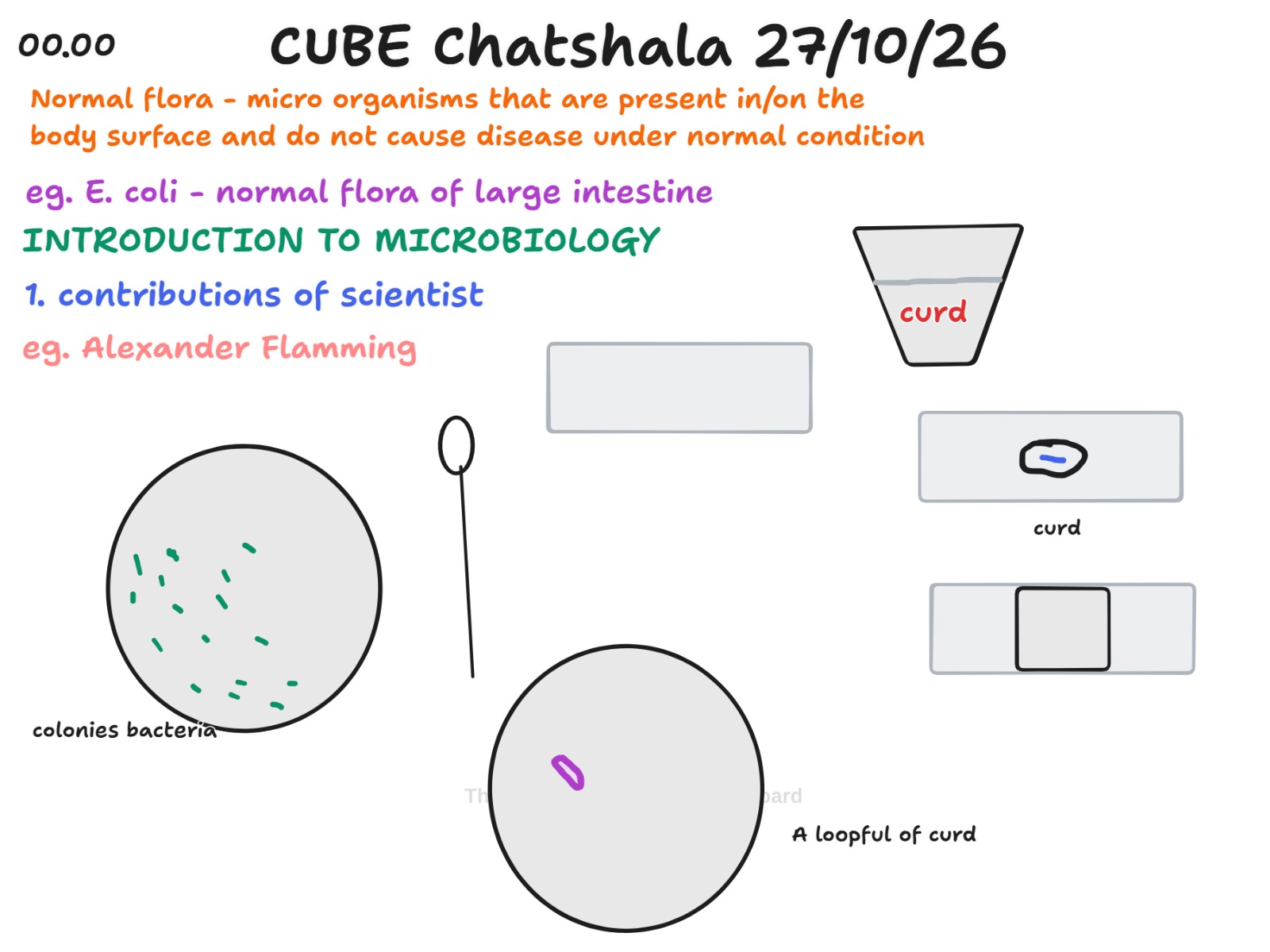
The whiteboard opened with the idea of normal flora, defined as the microorganisms that live in or on the surface of the body and do not cause disease under normal conditions. Escherichia coli in the large intestine was given as the standard example. Under the heading “Introduction to Microbiology”, the first topic listed was the contributions of scientists, with Alexander Fleming noted as the first name. These notes framed the evening’s central idea: microbes are not only agents of disease, and much of our understanding of them has been built by individual scientists working with simple tools.
The practical part of the discussion used curd as the working sample. The board showed a cup of curd, a loopful of curd placed on a plate, a plate with scattered bacterial colonies, an inoculation loop, and two slides. One slide carried a small curd smear with a blue line through it, representing a drop of methylene blue. The other showed a square coverslip placed over the smear. Sailekshmi’s slide-preparation notes described the same sequence: collect a tiny drop of curd or whey, spread it thinly on a clean slide, add the stain, and lower the coverslip at an angle so that air bubbles are not trapped. Aarya Takke then shared a step-by-step guide for Gram staining of Lactobacillus from curd. It covers smear preparation (a clean slide, transfer of the sample, air drying, heat fixing), the staining sequence of crystal violet, iodine, alcohol decolourisation, and safranin, and finally observation under oil immersion at 1000x. Its stated finding was that Lactobacillus is Gram-positive and rod-shaped.
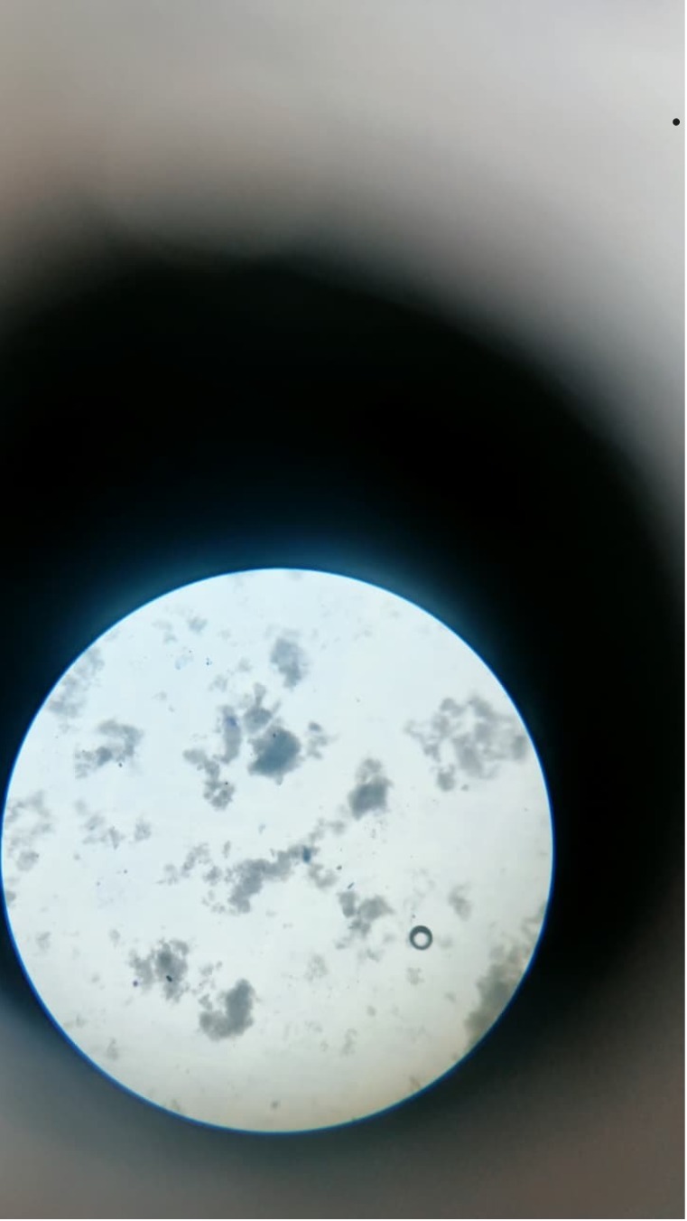
A microscope photograph of the curd preparation was also shared. It shows a bright circular field with a bluish-grey background and many irregular grey clumps. A small ring, most likely an air bubble, is visible near the lower right. The picture prompted a useful reminder that what a beginner sees through the eyepiece does not always match the neat purple rods drawn in a textbook diagram.
Rechel Tirkey contributed a description of the inoculation loop. As she explained it, the tool has a handle, a metal shaft, and a wire attached to the shaft that ends in either a round loop or a straight needle. This linked the whiteboard drawing of the loop to the actual instrument used to pick up a sample and streak it on a plate.
Later in the session, Rechel was appreciated for speaking to Om Gupta about the Nobel Prize, which brought the theme of scientists’ contributions back into the conversation. Sailekshmi shared the Royal Society profile of Venkatraman Ramakrishnan, a Nobel Prize-winning biologist known for determining the atomic structure of the ribosome, the cellular site where proteins are made. A research article on lactic acid bacteria isolated from sorghum silage in western Algeria was also shared as a reference. Its abstract reports that Lactobacillus made up the largest share of the 27 strains retained, followed by Weissella, Lactococcus, and Leuconostoc. This is a helpful reminder that lactic acid bacteria form a varied community and are not a single organism.
 Provocative Questions
Q1. Arunan MC asked what real learning is. If a student can recite the Gram staining steps perfectly but has never seen a stained smear, has that student really learned microbiology?
Q2. Normal flora are defined as microbes that do not cause disease under normal conditions. What exactly counts as normal, and what happens to the status of E. coli when it moves out of the large intestine?
Q3. Curd is made by bacteria, yet we eat it every day. Should we think of the bacteria in curd as normal flora, as food organisms, or as something in between?
Q4. The whiteboard shows both a stained smear on a slide and bacterial colonies on a plate. What different questions can each method answer about the same curd sample?
Q5. In the microscope photograph, the field is full of grey clumps. How can a beginner decide whether a clump is a cluster of bacteria, a particle of milk protein, or just debris?
Q6. A methylene blue smear and a Gram stain can both be done on curd. What does the Gram stain tell us that a simple stain cannot?
Q7. Alexander Fleming’s discovery began with an accidental observation. How much of scientific progress depends on carefully planned experiments, and how much on a prepared mind noticing something unexpected?
Q8. Why does the inoculation loop have a wire that ends in a small ring, and why is it heated before and after use?
Q9. The sorghum silage study found several different genera of lactic acid bacteria in one environment. If curd at home is made differently in each household, would we expect a different microbial community in each?
 What I Have Learned
The first thing I learned is that microbiology becomes much clearer when it begins with something familiar. Starting from a cup of curd made the idea of normal flora and beneficial bacteria feel close to everyday life, instead of something that only happens in a laboratory.
I also learned that the path from sample to observation has a definite order, and each step has a reason. A clean slide, a thin smear, air drying, and heat fixing prepare the cells so that they stay in place. The Gram staining steps then separate bacteria into two groups on the basis of their cell walls. Skipping or rushing any step changes what we see.
Rechel’s explanation of the inoculation loop taught me to look at the tool, and not only at the specimen. Knowing the parts of a loop, with its handle, shaft, and wire tip, makes it easier to understand how a sample is picked up and spread without contaminating it.
The microscope photograph showed me how different real observation is from a diagram. Textbook images show clean, clearly separated rods, while a real field can be crowded and unclear. Learning to read such a field honestly is a skill that comes only with practice.
Finally, the question about real learning stayed with me. Learning appeared to happen when someone tried a procedure, saw an unclear result, explained it to a peer, and corrected it together. The sessions work because people are willing to speak, listen,en and revise.
 TINKE Moments (This I Never Knew Earlier)
The moments below are drawn from what was recorded on the chat, the whiteboard,ard and the shared images.
-
The structure of an inoculation loop. Rechel Tirkey described the tool as a handle, a metal shaft, and a wire ending in either a round loop or a straight needle. For many of us, this was the first time the instrument was broken into its parts, and it explained why the loop in the whiteboard drawing looks the way it does.
-
Normal flora is a condition-based idea. The definition on the board carries the phrase “under normal conditions”. The learning here is that a microbe is not simply good or bad. Its role depends on where it is and on the state of the host.
-
Curd has a bacterial community, not one species. The sorghum silage reference showed several genera of lactic acid bacteria living together. This changed the way I think about the single word Lactobacillus used in the staining chart.
-
A real field of view is messy. The microscope photograph, with its grey clumps and a small bubble, showed that observation requires interpretation. A smear that looks unclear is still a result, and it teaches us how to improve the next preparation.
-
Scientific contribution can be very small in scale and very large in effect. Ramakrishnan’s work on the ribosome, a structure far too small to see directly, and Fleming’s observation on a plate both showed how careful looking can change medicine and biology.
 Gaps and Misconceptions
Gaps
Because the session relied on a chat log, a whiteboard, and shared images and not on a full record of what was said, some points were left open. The whiteboard lists Alexander Fleming under contributions of scientists, but the notes do not record what his contribution was. The Nobel Prize conversation involving Rechel and Om Gupta is mentioned in the chat, but its content was not written down.
The microscope photograph was shared without a note on the stain used, the magnification, or the objective lens. Without these details,ils it is hard to say what the grey clumps actually are, or whether the preparation was a simple methylene blue stain or a Gram stain.
The session also did not clearly connect the two methods drawn on the board. The plate with colonies and the slide with a smear answer different questions, but the link between them (growing bacteria first, and then staining a colony) was not spelled out.
Misconceptions to Clarify
-
Fleming’s surname is spelled Fleming, with one m. The whiteboard spelling was a slip that is worth correcting before the notes are shared.
-
A methylene blue smear is not a Gram stain. Methylene blue is a simple stain that shows shape and arrangement, while the Gram stain uses crystal violet, iodine, alcohol, and safranin and tells us about the cell wall. The two were shown close together in the shared material and could easily be confused.
-
Grey clumps in a curd smear are not necessarily bacteria. Curd contains milk proteins that form irregular masses and take up stain, and bubbles and debris can also appear in the field. Bacteria in curd are very small and need high power to be seen as individual cells.
-
Lactobacillus is not the only organism in curd. The infographic describes only Lactobacillus, but curd usually contains a mixture of lactic acid bacteria, and the sorghum silage study itself reported several genera.
-
The infographic describes Lactobacillus as anaerobic. This is a simplification, since many species are better described as aerotolerant, meaning that they can tolerate oxygen but do not use it for growth.
-
Normal flora does not mean harmless in every location. E. coli is a normal inhabitant of the large intestine, yet it can cause infection if it reaches other sites such as the urinary tract.
-
Curd bacteria are not strictly normal flora of the body. They are fermentation organisms taken in with food, and their role is closer to that of probiotics, which should be kept distinct from the resident flora described on the board.
 Photographs during Chatshaala

 Referance