 CUBE Chatshaala - Discussion Summary
Today’s ChatShaala session, held on 19th August 2026, opened with a warm welcome to two new participants joining the community for the first time. Shraddha, a second-year Zoology student from Elphinstone College with a minor in Biotechnology, and Rutuj Rajendra Nachankar, also a second-year Zoology student from the same institution with a minor in Botany, introduced themselves at the start of the meeting, adding fresh perspectives to the group’s ongoing conversations around microbiology.
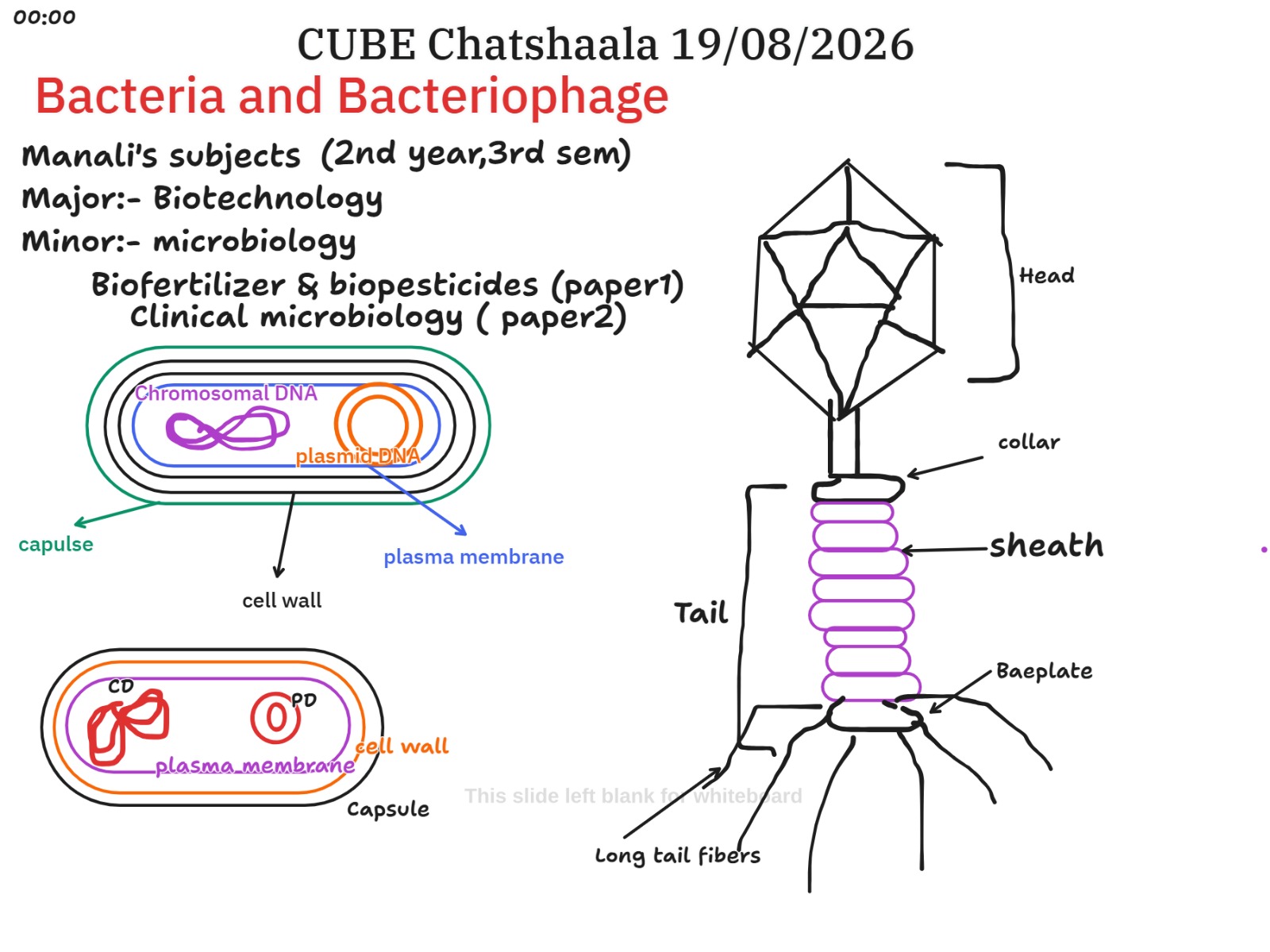
The core of the session revolved around the structural anatomy of bacterial cells and bacteriophages, which had been sketched out on the whiteboard as a visual anchor for the discussion. Manali, whose academic background spans a major in Biotechnology and a minor in Microbiology, along with coursework in biofertilizers, biopesticides, and clinical microbiology, contributed diagrams illustrating the bacterial cell in cross-section. These sketches labelled the key structural features of a bacterium, including the outer capsule, the cell wall, the plasma membrane, and the two distinct forms of genetic material found within, namely the chromosomal DNA and the smaller circular plasmid DNA. A parallel diagram of a bacteriophage broke down its architecture into recognisable components, from the icosahedral head down through the collar, the contractile sheath, the tail, the baseplate, and the long tail fibres that anchor the virus to its bacterial host during infection.
From there, the conversation moved naturally into a detailed exploration of how penicillin works against bacteria and why some bacterial populations manage to survive exposure to it. Aarya led this portion of the discussion, explaining that bacteria lacking a penicillin resistance gene are killed when exposed to the antibiotic, whereas those carrying the gene survive because it codes for an enzyme that neutralises the drug’s effect. She described the underlying mechanism in some depth: penicillin interferes with the synthesis of the bacterial cell wall, and once that protective barrier is compromised, excess material floods into the cell. The resulting imbalance in osmotic pressure causes the cell to swell and eventually burst. Sailekshmi built on this by clarifying that penicillin specifically blocks the enzymes bacteria rely on to build and repair their cell walls, which is what leaves the cell vulnerable to rupture. Rutuj then offered a concise synthesis of the mechanism, describing penicillin as a beta-lactam antibiotic that disrupts the penta-peptide cross-links within the cell wall, leaving the structure unable to withstand internal osmotic pressure.
The session also touched on a more fundamental distinction that often gets glossed over in casual conversation: the difference between viruses and bacteria as biological entities. Unlike bacteria, viruses cannot be cultured independently on standard growth media because they lack the cellular machinery to replicate on their own and are entirely dependent on a host cell to reproduce. This dependency is one of the clearest lines separating the two, alongside the difference in ribosome structure: prokaryotic bacteria carry 70S ribosomes, while eukaryotic cells, including human cells, carry the larger 80S variety. This ribosomal distinction came up when Rutuj shared the comparison and then caught and corrected a small error in the initial statement, a moment that Niharika met with the reassuring observation that mistakes are simply part of the learning process.
 Provocative Questions
-
If penicillin only affects bacteria by targeting cell wall synthesis, why does this mechanism have no equivalent vulnerability to exploit in human cells, and what does that tell us about the evolutionary divergence between prokaryotic and eukaryotic cell architecture?
-
Given that plasmid DNA can carry resistance genes independently of chromosomal DNA, how significant a role might horizontal gene transfer between bacteria play in the rapid spread of antibiotic resistance within a population?
-
Bacteriophages rely on remarkably intricate structures, the contractile sheath, the baseplate, the tail fibres, purely to inject genetic material into a host. What evolutionary pressures might have shaped such mechanical precision in something as structurally minimal as a virus?
-
If viruses cannot be grown outside a host cell, how do researchers study bacteriophage biology and structure in a laboratory setting, and what does this constraint reveal about the broader challenge of studying obligate parasites?
-
Why might a bacterium’s capsule, an outer layer distinct from the cell wall, offer an additional survival advantage, and how does this feature interact with a host immune system during infection?
 What I Have Learned
Working through this session reaffirmed how much clarity comes from pairing a structural diagram with a functional explanation. Seeing the bacterial cell broken into its component layers, capsule, cell wall, plasma membrane, and then tracing where the chromosomal and plasmid DNA sit within that architecture, made the abstract idea of antibiotic resistance feel far more concrete. It is one thing to know that resistance genes exist; it is another to see exactly where that genetic material resides and to understand how a plasmid can carry a resistance trait independently of the cell’s main chromosome.
The breakdown of the bacteriophage structure was equally valuable. The distinction between the head, collar, sheath, tail, and baseplate is not merely anatomical trivia; it maps directly onto how the virus mechanically punctures a bacterial cell wall and delivers its genetic payload. That level of mechanical sophistication in something so much simpler than a bacterial cell is a genuine reminder of how much complexity can exist within very small biological systems.
The penicillin discussion tied these threads together nicely. Understanding that the antibiotic disrupts cell wall synthesis by blocking the enzymes responsible for forming penta-peptide cross-links, and that this disruption leads to osmotic rupture, gives the whole concept of “bacteria dying from penicillin” a mechanistic backbone rather than leaving it as a vague fact to memorise. It was also useful to be reminded that resistance is not simply a matter of a bacterium being “immune” in some abstract sense, but that it comes down to a specific enzyme encoded by a specific gene actively protecting the cell wall from the drug’s action.
 TINKE Moments (This I Never Knew Earlier)
The clearest TINKE moment of the session came from Rutuj’s initial statement on ribosome types, where the eukaryotic and prokaryotic classifications were briefly mixed up before being corrected in real time. This is a genuinely common point of confusion, since the terminology (70S versus 80S) is easy to transpose if not anchored firmly to which cell type each belongs to. The self-correction, followed by Niharika’s supportive response that learning through mistakes is a natural and welcome part of the process, captured the collaborative and low-pressure spirit that ChatShaala aims to cultivate. It reinforced that the space is designed for participants to think aloud, test their understanding, and revise it openly without any sense of judgement.
A second, more implicit TINKE moment emerged from the layered explanation of the penicillin mechanism itself. As the explanation moved from Aarya’s initial framing, through Sailekshmi’s clarification, to Rutuj’s more technical synthesis involving beta-lactam structure and penta-peptide cross-links, the discussion effectively built understanding in stages. This progression suggests that the initial framing of “penicillin kills bacteria without the resistance gene” was accurate but incomplete, and it took successive contributions to arrive at a fuller mechanistic picture, a useful illustration of how scientific explanations often deepen through group dialogue rather than arriving fully formed from a single source.
 Gaps and Misconceptions
The session’s discussion, while rich in structural and mechanistic detail, did not explore how antibiotic resistance genes actually spread between bacterial populations, whether through conjugation, transformation, or transduction, which would have connected naturally to the plasmid DNA already diagrammed on the whiteboard. Similarly, while the bacteriophage structure was described in detail, the session did not touch on the broader life cycle distinction between lytic and lysogenic phages, which would help explain why some phages destroy their host immediately while others integrate into the bacterial genome. The connection between the two reference materials on viruses, the general virus-versus-bacteria comparison and the coronavirus-specific material, and the more bacteriophage-focused whiteboard content also remained somewhat underexplored, leaving an opportunity to discuss how bacteriophages differ from viruses that infect human cells, such as coronaviruses, in terms of structure and host range.
No major misconceptions persisted uncorrected by the end of the session. The one point of confusion that did surface, the initial mix-up between 70S and 80S ribosomes across prokaryotic and eukaryotic cells, was identified and corrected within the same exchange, which is precisely the kind of self-correcting dialogue this format is designed to encourage. It is worth noting, for future reference, that this particular ribosome mix-up is a recurring stumbling block in introductory microbiology discussions generally, and may be worth revisiting briefly in a future session to reinforce the distinction.
 Photographs during Chatshaala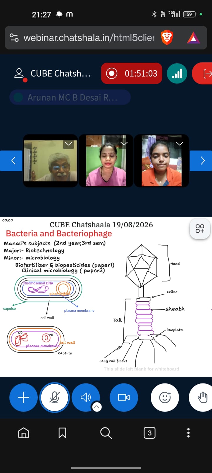

 Referance