All thanks to CUBE Chatshaala sessions, i came up with this great idea of making DIY Phone Microscope. During this Lockdown period, we (CUBist) have made a lab at Home named ( CUBE HOME LAB) and trying to Explore new ways of Learning.
Similarly i have tried to make a Phone Microscope that would allow me to explore tiny objects.
I would like to give some Examples related to our CUBE HOME LAB Work.
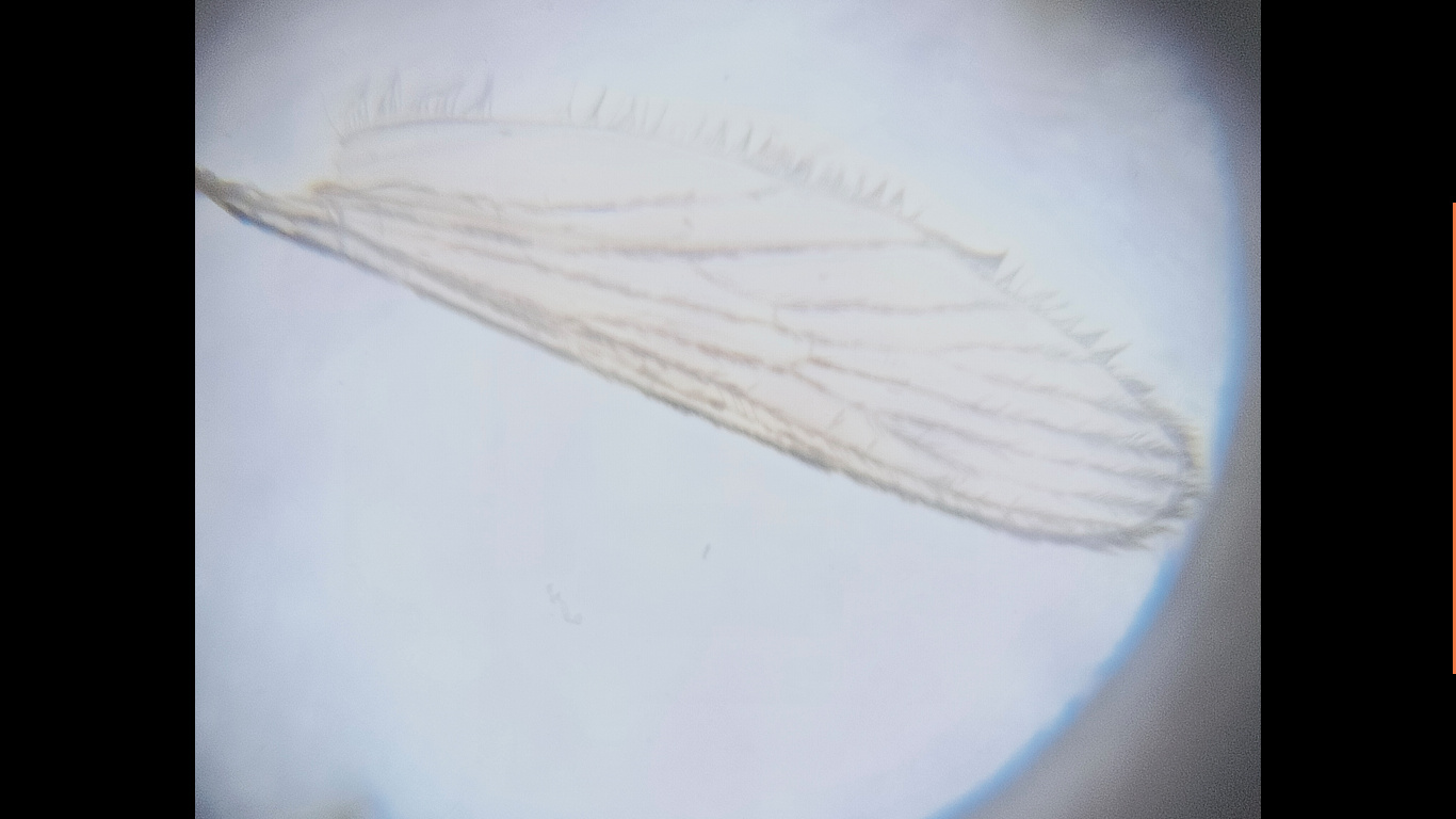
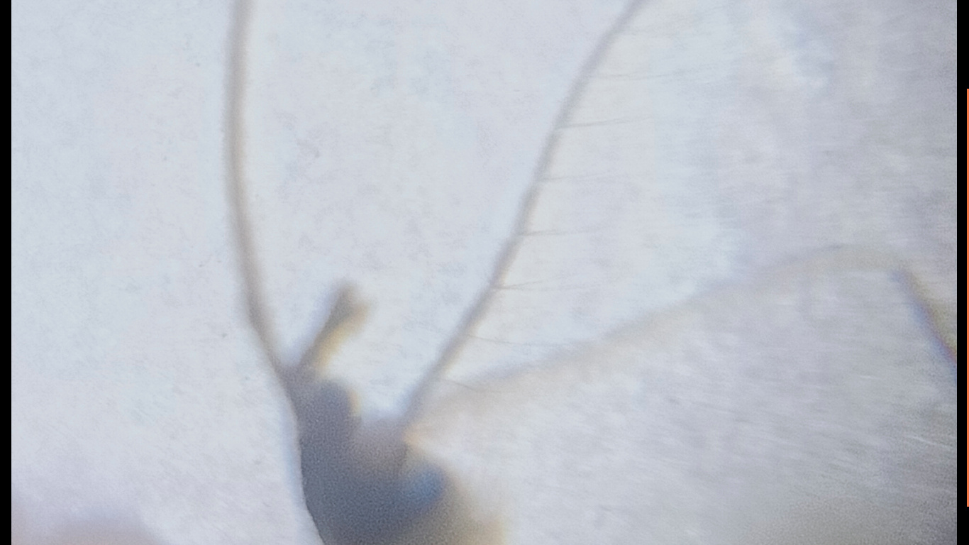
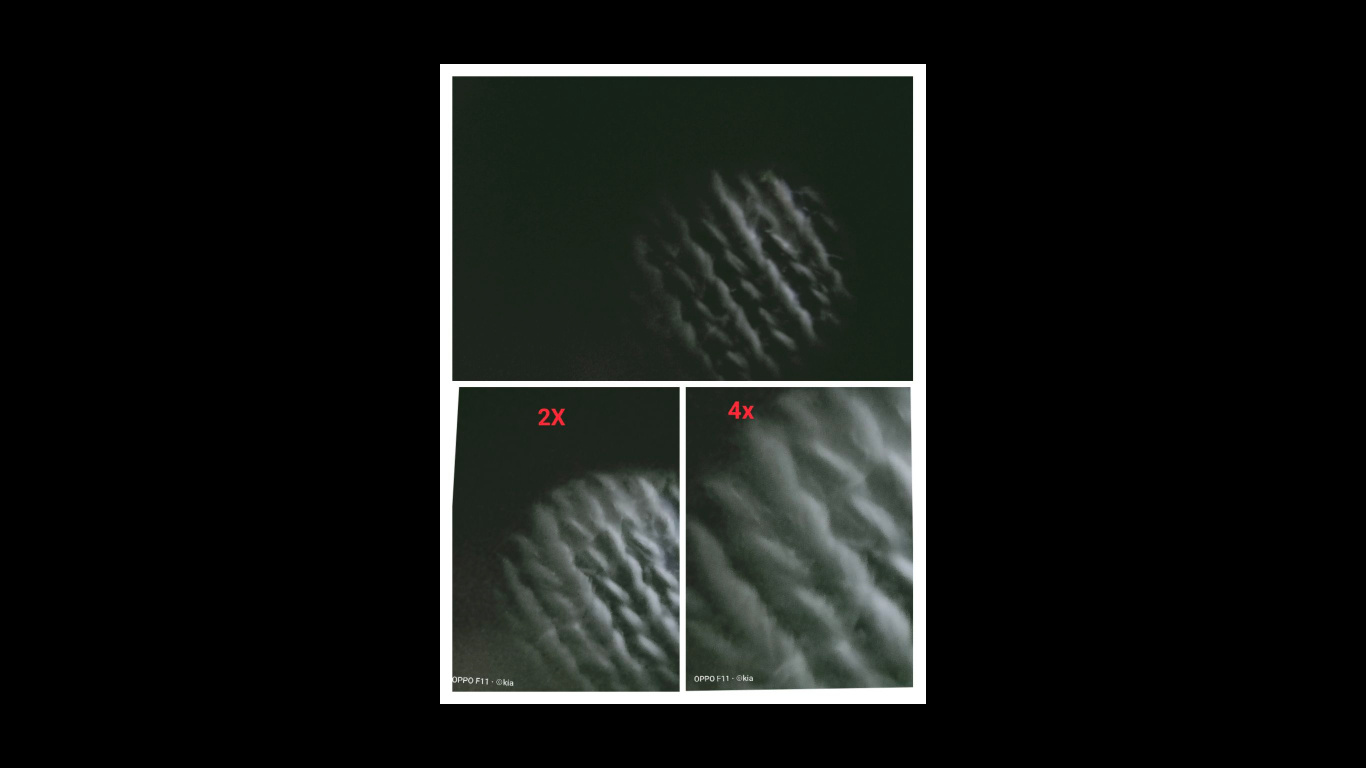
To see Two Breaks in Coastal vein, an incomplete sub-coastal vein and an Anal cell which are present in the wing of a Fruit Fly ( Characteristics features of Drosophilidae family)
To see tiny living creatures living in a drop of puddle water using Phone Microscope.
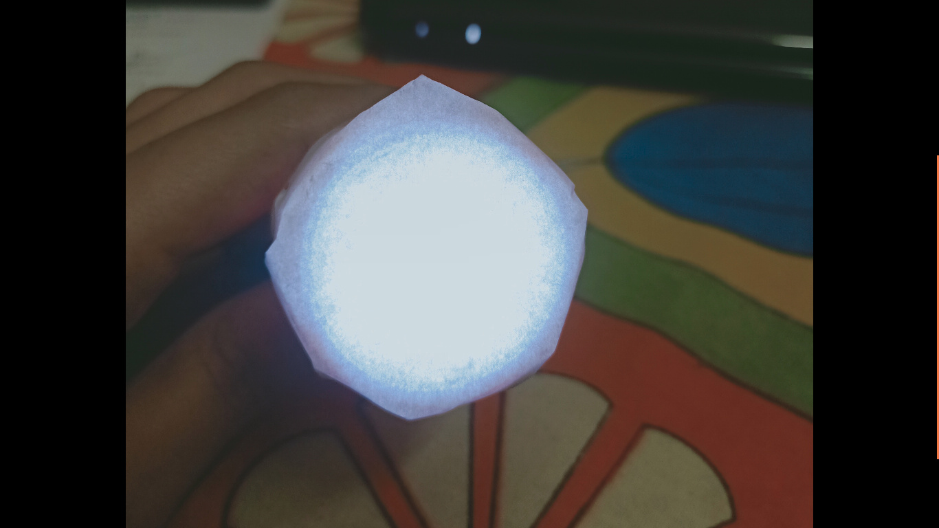
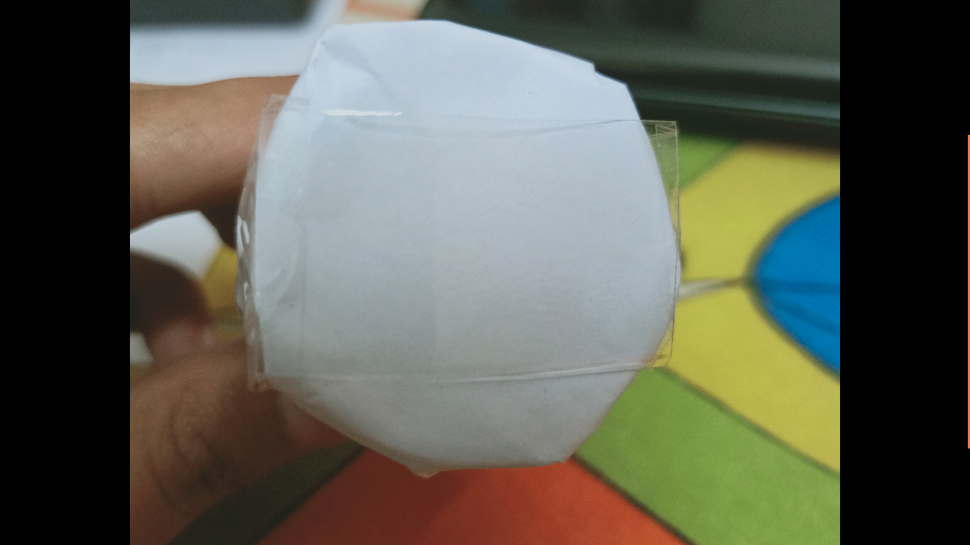
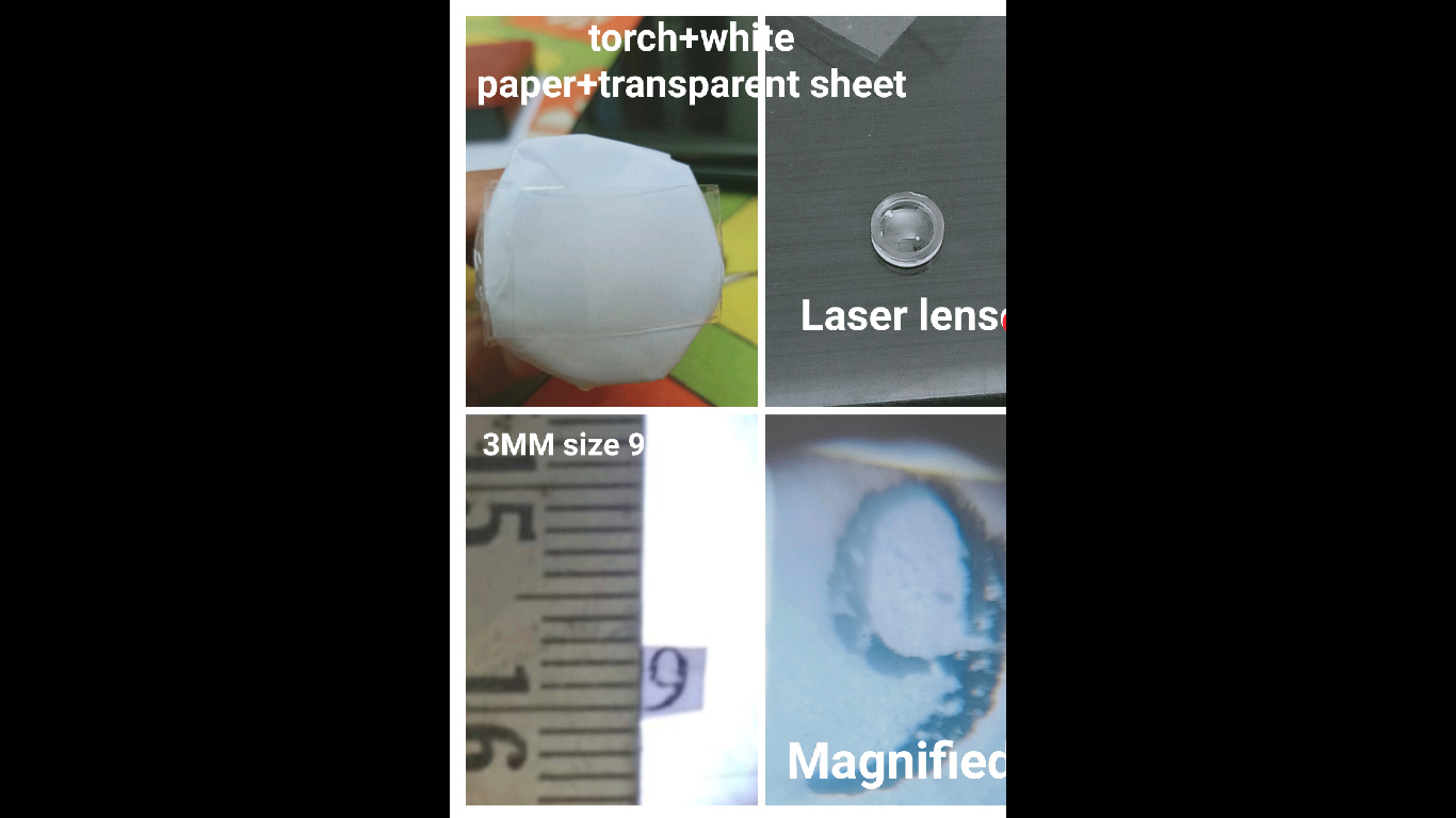
Requirements: The things i have used to make Phone Microscope were available in my house.
A small white light torch
A piece of plane paper
A transparent plastic sheet ( i have used transparent file cover)
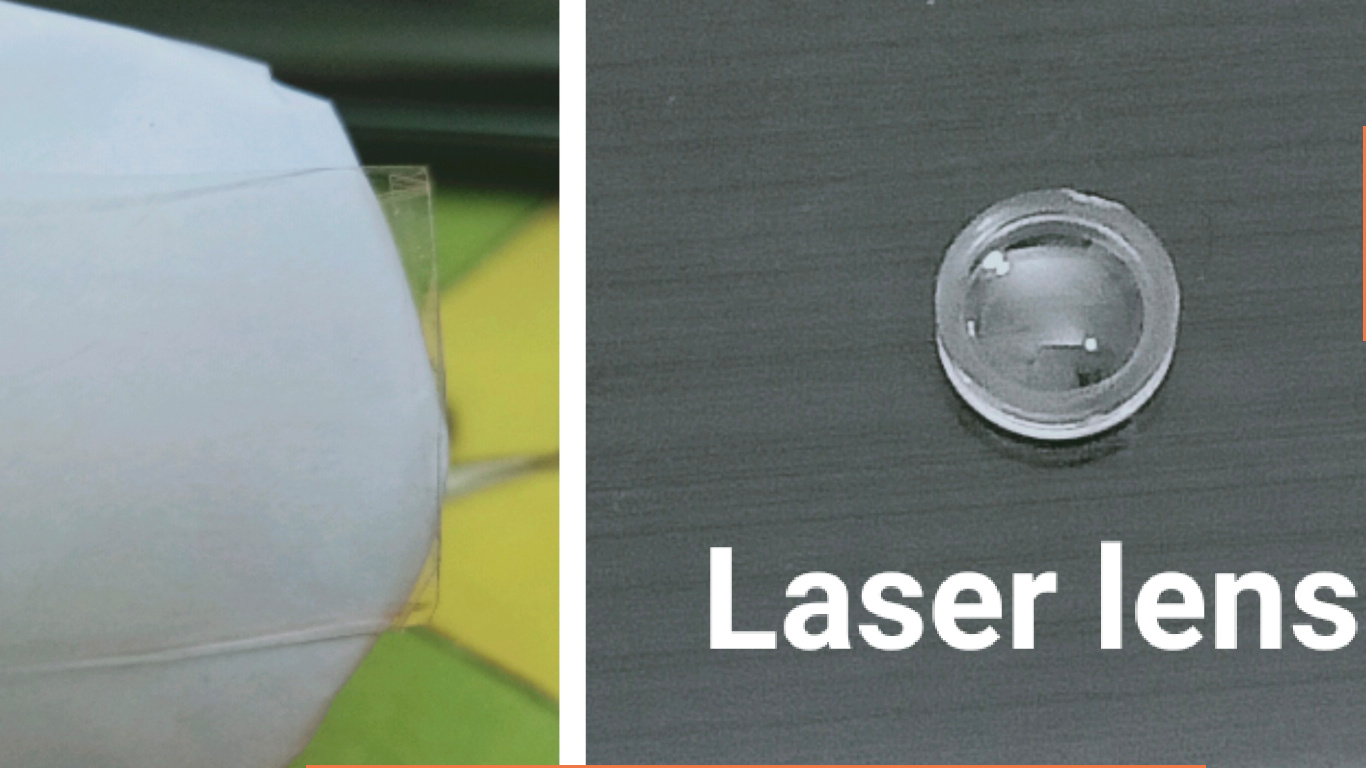
Laser Pointer ( which is also available in the market in 20 to 30rs.)
Physicists and Makers are becoming extremely interested in biology. Eg photosynthesis and the plethora of molecules used by plants to optimise photovoltaic effect such that minimal damage is caused by UV while optimal efficiency is maintained. A trick that can be applied to solar cells.10.1126@science.aba6630.pdf (823.8 KB) 10.1126@science.abc8063.pdf (239.1 KB)
help !!! 4
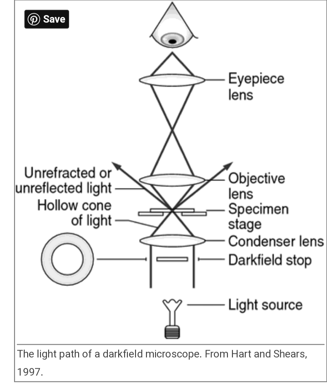
This is a ray diagram of a compound microscope, and all of us at Cube Chatshaala! are trying to understand how a microscope processes an image of an object. Could anyone help me explain how the image is formed in the mobile phone camera?? Probably ray diagram of a mobile phone camera.
Thank you!!! @jtd@ Asmita@Jaikishan and others
@saswathy679 has been working on fruit flies. To identify the fruitflies she needed to take a close look at their wings. She tried doing it with her mobile phone camera and torchlight. She has been reasonably successful, but to get better clarity, we started exploring more options.
@Farhan and @GN suggested last week to not use digital zoom on her phone, to take a close enough picture directly and to zoom it later for better results.
@saswathy679, we are waiting for the results. You can start by posting the before and after pictures here, and we will continue the discussion.
Also, I think this can be a wonderful exploration for other investigations at home labs as well. For example, @saida786110 wanted to check the rotifers using a mobile camera. There was a similar exploration here tinkering a lens from the DVD by @singh (See here).
It’s here!
Adjust the video resolution to 1080p for clarity.
Fungus in its own niche captured via mobile microscopy
Growing on an empty stalk of a custard apple.
Are these conidia? What do we call the white filament-like structures?
Taken via a convex lens obtained from laser light.
No digital zooming was done.
This is beautiful. It is tough to imagine that ugly looking fungus can appear so good when magnified. What are the typical features by which we distinguish fungus?
Mushrooms also have a similar structure. Do their heads also have spores? Or am I completely off?
True! And to add to the curiosity, perhaps this is just one type of fungi out of around 1,44,000 species (Britannica) including invasive as well as non-invasive ones!
Fungi typically have these (as seen in the video) filamentous (hair-like) projections called sporangium (the stalk) and the bulb houses the spores. These are of varying colours (white, green, black), depending upon the species and it’ll visible only when there is sufficient growth on the substrate. But again, not all fungi show these structures.
You can try to invite them to your place!
Just keep a slice of bread or a piece of chapati near the dustbin/kitchen sink and sprinkle some water. Wait for 24-48hrs and it’ll be there but inorder to visualise them initially, you’ll have to look-closely in a bright outing.
No, they don’t have spores… The spores will be present in their heads [called Caps (pileus)]
A spore wrt. fungi is something like a reproductive body that when attached to a substrate (bread, chapati), will lead to the growth of that fungus.