 CUBE ChatShaala - Discussion Summary
Today’s ChatShaala session, held on August 5, 2026, wove together two threads that at first glance seem unrelated but actually complement each other rather nicely: the fine architecture of bacterial cells versus viruses, and a live fermentation experiment on curd-making. The group included Sailekshmi, Manali Bhujade, Aarya, Niharika, Kiran K., Himanshu Joshi, and Arunan (joining from B Desai Road, Mumbai).
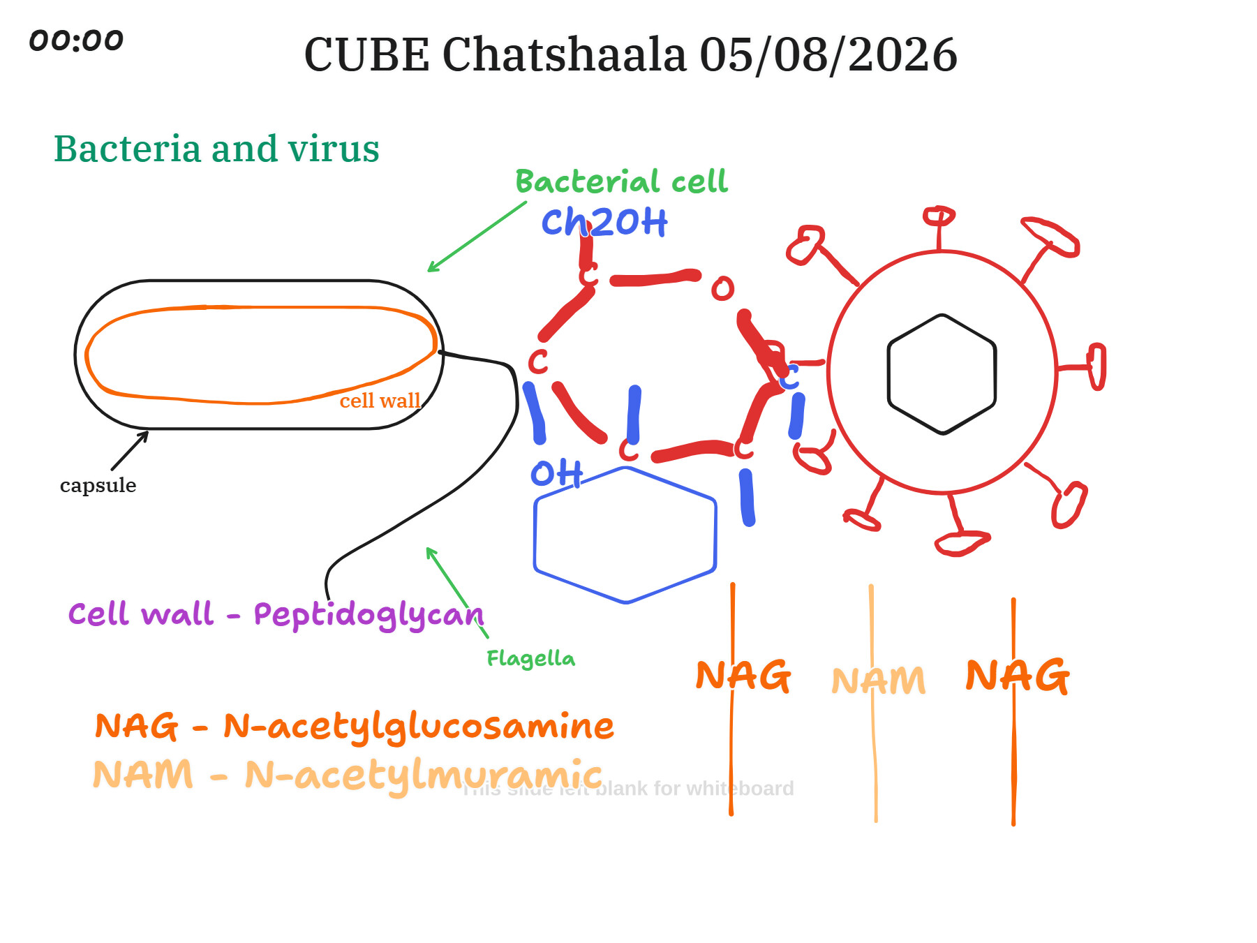
The session opened with a whiteboard comparison of bacterial and viral structure. The discussion traced the outer boundary of a bacterial cell from the outside in: the capsule, a gelatinous protective envelope; then the cell wall, whose main structural component is peptidoglycan (also called murein); and finally the cytoplasm, drawn as an oval enclosing the genetic material. The peptidoglycan itself was broken down into its two alternating sugar units, N-acetylglucosamine (NAG) and N-acetylmuramic acid (NAM), joined in a repeating chain that gives the wall its mesh-like, almost chain-link quality. This sugar backbone alternates in a β-(1,4) linkage, and each NAM unit carries a short chain of amino acids that cross-links to neighbouring strands, producing the three-dimensional mesh that gives the wall its strength. The flagella were also marked on the diagram, branching off the cell wall as the structure responsible for bacterial movement.
Alongside this, the virus was sketched as a much simpler, spiked sphere, a reminder of just how different viral and bacterial architecture really are. Where a bacterium is a self-sufficient cell with its own wall, membrane, and machinery, a virus is little more than genetic material wrapped in a protein coat, spikes and all, entirely dependent on a host cell to replicate. Sitting the two diagrams side by side made the contrast concrete in a way that reading about it separately often doesn’t.
The capsule discussion added an important nuance: this outer gelatinous layer is what gives certain bacteria their virulence, since capsulated bacteria can cause disease while non-capsulated ones generally cannot. Its slippery, negatively charged surface makes it harder for immune cells called phagocytes to grab onto and engulf the bacterium, which is part of why capsulated strains are more likely to evade the body’s defences. The capsule also guards against drying out, since it holds a fair amount of water, and it helps bacteria stick to host tissue and to each other, which is often the first step toward forming a biofilm.
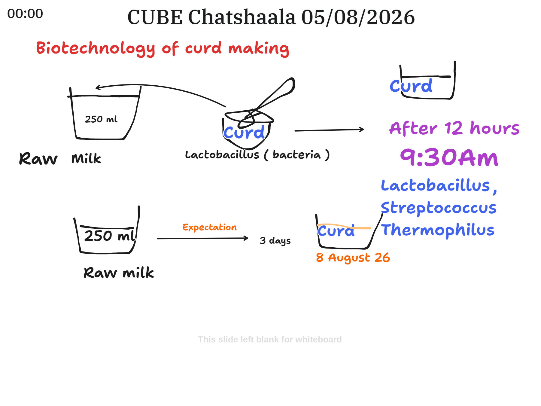
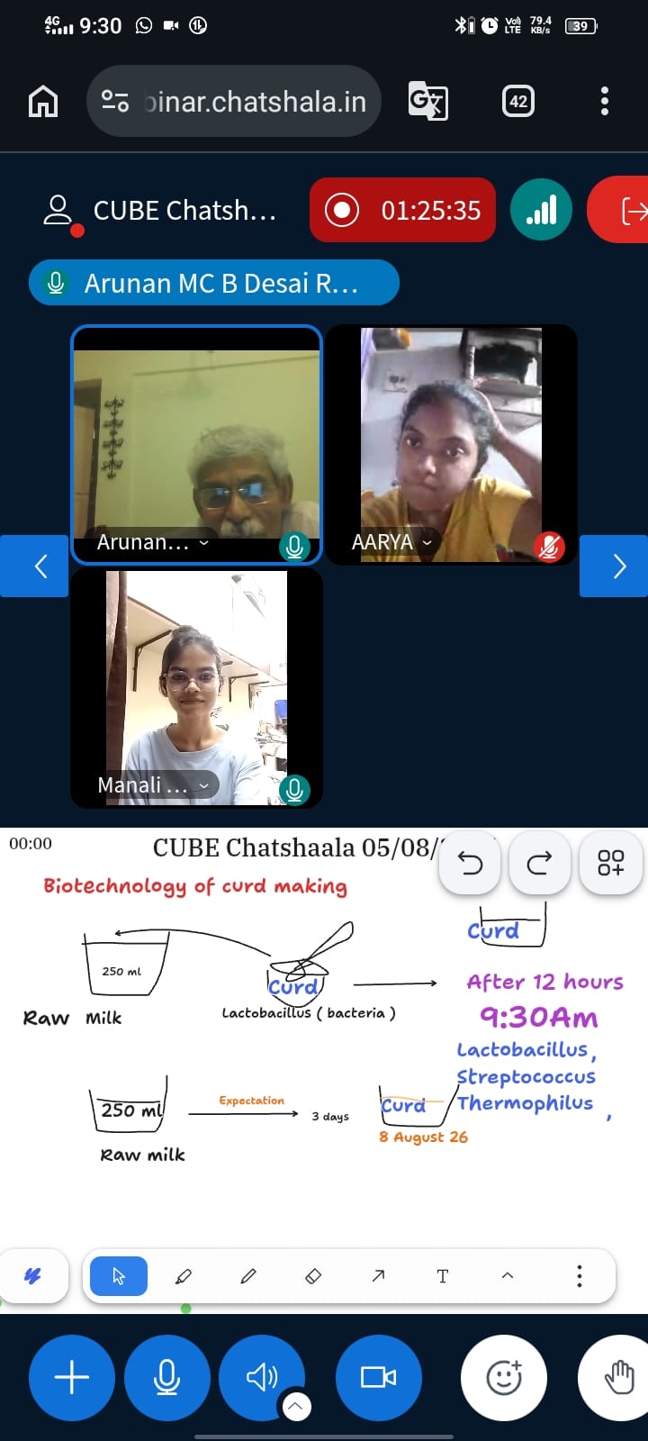
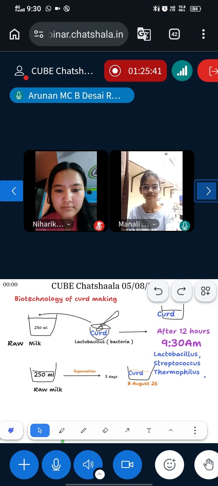
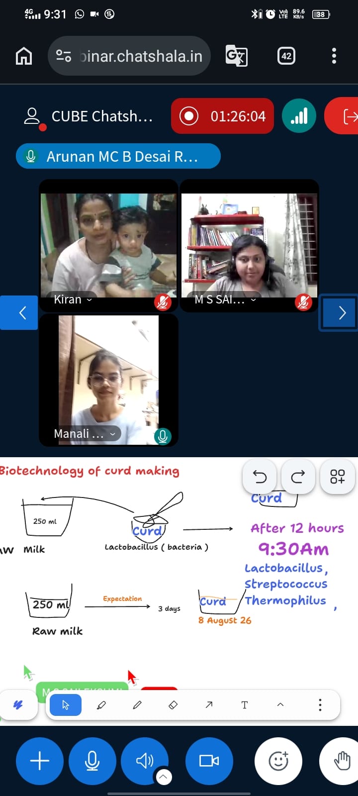
The second half of the session moved from structure to practice, with a hands-on look at the biotechnology behind curd-making. Two parallel setups were described on the whiteboard. In the first, 250 ml of raw milk was inoculated with a spoonful of curd containing live Lactobacillus, and within twelve hours (by 9:30 am the next morning) it had set into curd — with Lactobacillus and Streptococcus thermophilus specifically named as the working cultures. In the second setup, a fresh 250 ml batch of raw milk was left to set on its own, with an expectation of curdling within three days; this batch dated August 8, 2026. The comparison highlights a point that often gets glossed over in textbook descriptions of fermentation: culturing with an existing starter dramatically speeds up the process compared to relying on whatever bacteria happen to be present naturally in the milk, and the specific bacterial species involved make a measurable difference to both the timeline and, presumably, the taste and texture of the final product.
Taken together, the two parts of today’s session reinforced the same underlying idea from different angles: that bacteria are living, structurally complex organisms whose cell walls and surface features aren’t just decorative, but actively shape how they interact with their environment, whether that’s evading a host immune system or converting milk into curd.
 Provocative Questions
-
If peptidoglycan is what gives bacterial cell walls their strength, why do antibiotics like penicillin target this layer specifically, and what happens to a bacterium once that wall is compromised?
-
Viruses lack a cell wall or capsule altogether, relying instead on a simple protein coat. Does this structural minimalism make them more or less vulnerable than bacteria, and why?
-
Since capsulated bacteria are typically more pathogenic than non-capsulated ones, could the capsule alone be a viable target for future antibiotics or vaccines, sidestepping the need to kill the bacterium outright?
-
In the curd experiment, one setup was deliberately inoculated while the other was left to ferment naturally. What does the difference in setting time tell us about the role of starter cultures versus ambient/wild bacteria in fermentation?
-
Both Lactobacillus and Streptococcus thermophilus were involved in the faster-setting curd. What might each organism be contributing individually, and would using only one species change the outcome?
-
If a virus needs a host cell to replicate but a bacterium has all its own cellular machinery, what does that suggest about how each evolved, and which came first in evolutionary history?
 What I Have Learned
Today’s session really sharpened my understanding of just how deceptively simple the phrase “bacterial cell wall” is when you first hear it, and how much complexity sits underneath. Seeing the NAG-NAM chain drawn out sugar by sugar made the peptidoglycan structure click in a way that reading the term in a textbook never quite managed. I also came away with a clearer sense of why the capsule matters clinically and not just structurally — it’s not simply a protective layer; it’s actively involved in how a bacterium interacts with (and sometimes outsmarts) a host’s immune system.
The curd experiment was a nice grounding exercise after all that structural detail. It’s one thing to know that Lactobacillus ferments milk into curd; it’s another to watch two batches running in parallel and see, in real time, how much of a difference an inoculant makes to the timeline. The twelve-hour versus three-day contrast was a genuinely useful, tangible way to internalize what “culturing” actually means in practice.
 TINKE Moments (This I Never Knew Earlier)
-
The wall is not one thing, it’s a woven structure. I had a vague sense that bacterial cell walls were “made of peptidoglycan,” but I hadn’t explicitly registered that this means an alternating chain of two distinct sugar units (NAG and NAM), cross-linked by short peptide bridges. Naming the components individually made the structural logic of the wall explicit for the first time.
-
The capsule and the cell wall are not the same layer. Before today, I sometimes used “capsule” and “cell wall” somewhat interchangeably in casual thinking. The session made explicit that the capsule sits outside the cell wall as a separate, distinct structure with its own composition and its own specific function (namely, virulence and immune evasion), rather than being just another name for the wall itself.
-
Fermentation speed is a direct, visible readout of microbial activity. I understood in the abstract that starter cultures speed up fermentation, but running the inoculated and non-inoculated milk samples side by side made explicit just how large that difference can be — a matter of hours versus days — and turned an abstract fact into something I now know from direct comparison.
 Gaps and Misconceptions
-
The exact mechanism by which the capsule’s negative charge repels phagocytes wasn’t detailed at the electrochemical level during the session; this is worth a closer look for anyone wanting the full immunological picture.
-
The relative individual contributions of Lactobacillus versus Streptococcus thermophilus in the faster-setting curd batch weren’t separated. It would be a useful follow-up experiment to try single-culture inoculations to isolate each organism’s specific role in texture and setting time.
-
The session didn’t explicitly address why the naturally fermenting batch (no starter added) was expected to take three full days; is this due to a lower starting population of relevant bacteria in raw milk, competition from non-fermenting microbes, or slower environmental conditions? This causal mechanism remains a gap worth exploring in a future session.
-
The virus diagram, while useful for contrast, didn’t get into how a virus actually attaches to and enters a host cell a natural next step given today’s focus on bacterial surface structures.
 Photographs during Chatshaala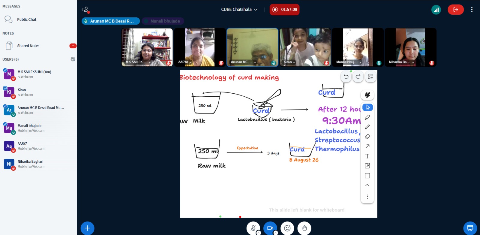
 Referance