 CUBE Chatshaala - Discussion Summary
Today’s CUBE ChatShaala session, held on 24th August 2026, centered around Niharika’s ongoing CUBE HomeLab work in Kathayatbara, Bageshwar, Uttarakhand, where she has been growing onion roots to study mitosis. The session brought together Sailekshmi, Manali Bhujade, Aarya Takke, Niharika Baghari, Kiran Kalakoti, Arunan MC, Kiran Varma, and Chitralekha, with the discussion moving fluidly between hands-on observation, hypothesis testing, and deeper conceptual biology.
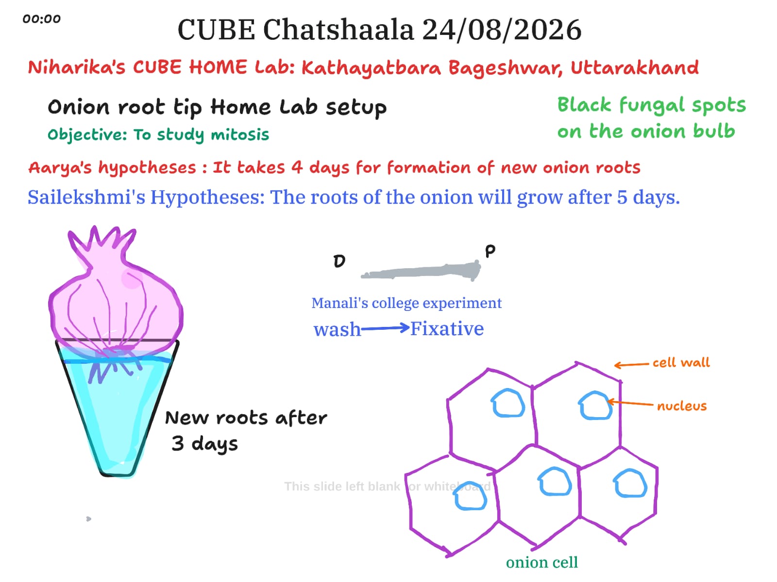
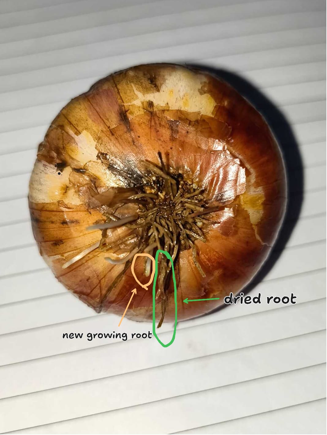
The core experiment involved placing an onion bulb over water to observe root tip growth, to eventually study mitotic cell division under the microscope. Interestingly, the group did not converge on a single prediction for how long root formation would take. Aarya hypothesised that new roots would take about four days to appear, while Sailekshmi proposed five days. The actual observation, as captured in Niharika’s photograph, showed new roots emerging within three days, alongside a clearly visible dried root from a previous growth cycle. This photograph became a valuable teaching moment, as Aarya asked Niharika how she was able to distinguish the newly grown root from the old one purely through observation, a simple but important question about how careful visual documentation supports scientific claims.
Arunan steered the conversation toward the “why” behind the HomeLab model itself, asking participants to reflect on the purpose of running these experiments at home rather than only in a formal lab setting. He also asked Niharika about the two HomeLabs she has set up in Uttarakhand and which specific experiments have begun at the Kathayatbara site. In the spirit of keeping the HomeLab approach genuinely participatory, Arunan encouraged everyone in the group to pick up an onion from their own kitchen, observe it, and share a photograph, turning the exercise into a distributed, hands-on citizen science activity rather than something observed only by one person.
The participants also worked through Manali’s college-level protocol for preparing onion root tips for microscopy, which follows a wash-to-fixative sequence. This opened up a discussion on what a fixative solution actually does. Kiran Varma explained it simply as a chemical used to preserve cells and tissues by halting decomposition, while Sailekshmi added that fixatives also harden the sample, making it suitable for sectioning and viewing under a microscope, with 10% neutral buffered formalin cited as a common example.
From there, the conversation moved into more advanced territory. Arunan introduced the idea of the onion membrane as a biosensor material, describing how the inner epidermis of the onion bulb scale can serve as a natural support structure for immobilising the enzyme Glucose Oxidase, which is used in biosensors designed to measure dissolved oxygen. Sailekshmi translated this into more accessible terms, noting that onion membranes are an appealing alternative to synthetic membranes because they are inexpensive, naturally available, transparent, and environmentally friendly, making them useful for anchoring enzymes in low-cost biosensor designs.
The participants then revisited foundational definitions to make sure everyone was working from the same base understanding. Aarya defined tissue as a group of cells with similar structures that function together as a unit, along with the intercellular matrix that fills the spaces between them. Kiran offered a complementary definition, describing tissue as a group of similar cells performing a specific function, and identifying the cell as the smallest basic unit of life. Arunan closed out the conceptual portion of the discussion with an explanation of the plant cell vacuole, describing it as a large fluid-filled compartment bound by the tonoplast membrane that can occupy up to 90 percent of a mature plant cell’s volume, storing water, nutrients, and waste products while maintaining turgor pressure that keeps the cell structurally rigid.
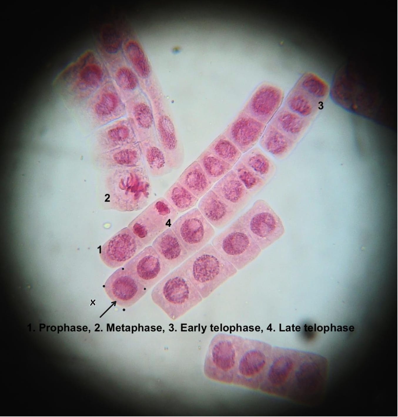
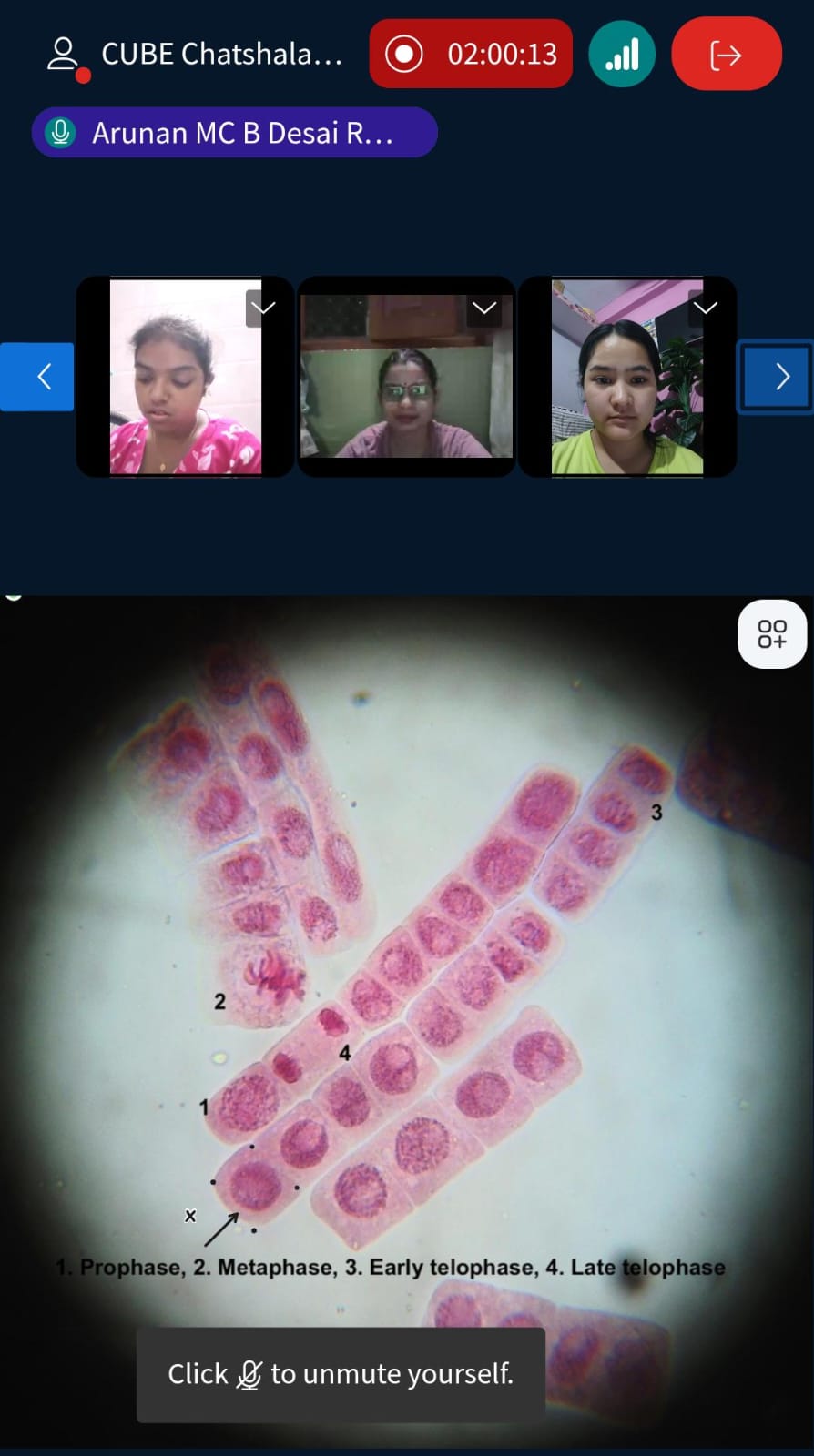
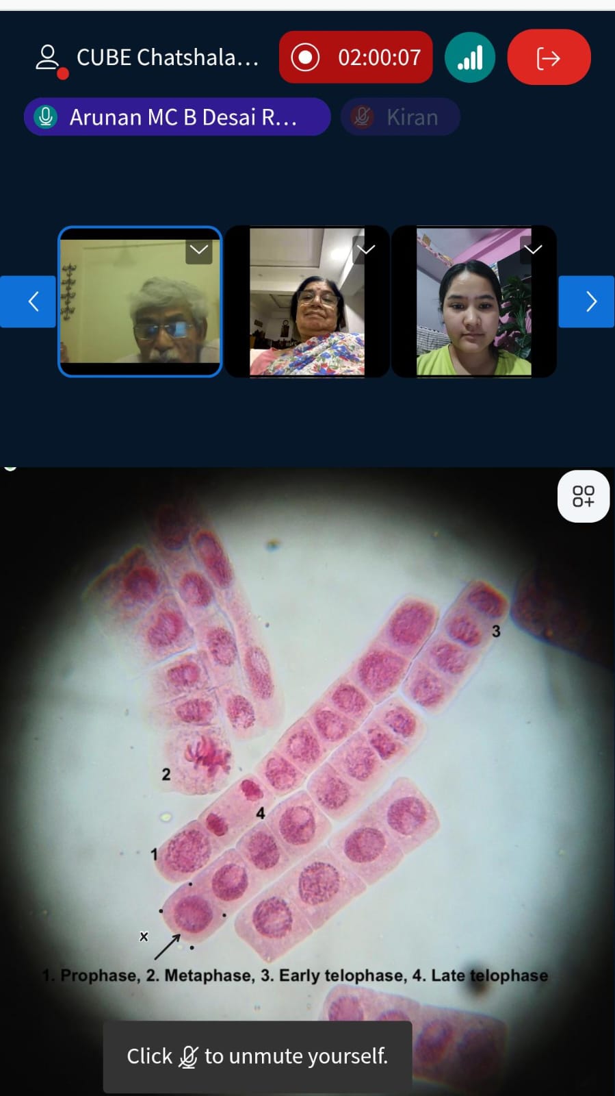
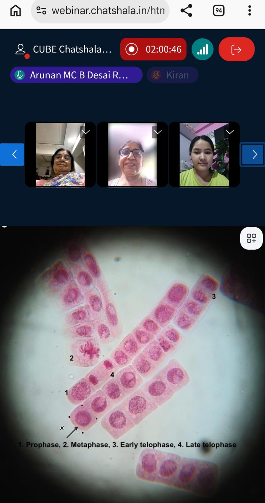
The microscope images shared during the session showed onion root tip cells at different stages of mitosis, clearly labelled as prophase, metaphase, early telophase, and late telophase, giving the group a direct visual anchor for the theoretical discussion around cell division. Niharika also shared a short video resource on onion structure to supplement the visual learning for the participants.
 Provocative Questions
-
If a researcher only observes an onion once, without a prior baseline photograph, how reliable is their claim about which root is newly grown versus older growth?
-
Given that Aarya predicted four days and Sailekshmi predicted five days, but the actual growth appeared at three days, what environmental or biological factors might explain such variation in root emergence timelines across different setups?
-
Why might running this experiment as a distributed HomeLab activity, with many participants observing their own onions, produce more reliable scientific insight than a single centralised observation?
-
What specific properties of a fixative solution allow it to both halt cellular decomposition and prepare tissue for microscopic sectioning at the same time?
-
Why would a naturally occurring membrane, such as onion epidermis, be considered advantageous over synthetic membranes in enzyme-based biosensor design, beyond simply being cheaper?
-
If the vacuole can occupy up to 90 percent of a plant cell’s volume, what does this suggest about how plant cells achieve structural support compared to animal cells, which lack this feature?
-
Looking at the labelled mitosis image, what visual cues distinguish early telophase from late telophase, and why does this distinction matter for understanding the pace of cell division?
 What I Have Learned
This session reinforced how much can be learned from a single, humble onion when it is examined carefully and from multiple angles. The differing hypotheses around root growth timing were a useful reminder that even a simple prediction, when tested against real observation, can reveal how much natural variability exists in biological systems, and that being wrong about the exact number of days is far less important than the discipline of predicting, observing, and comparing.
I also came away with a clearer appreciation for how the same everyday material, in this case the onion, can serve entirely different scientific purposes depending on the question being asked. On one hand, the root tip becomes a window into mitosis and cell division. On the other, the inner membrane becomes a functional material in biosensor technology. This dual use illustrates how citizen science and applied research are not as far apart as they might initially seem, and how a low-cost home experiment can echo principles used in more sophisticated laboratory applications.
The discussion around fixatives and the wash-to-fixative protocol also helped connect the practical steps of sample preparation to the underlying purpose behind each step, rather than treating them as a rote procedure. Understanding that fixation both preserves and hardens tissue gives the process more scientific meaning, rather than it being just a step to move past on the way to viewing slides under a microscope.
 TINKE Moments (This I Never Knew Earlier)
The most significant TINKE moment of the session emerged from Aarya’s question to Niharika about identifying the newly grown root purely through observation. It became clear through this exchange that visual identification of new growth without a documented baseline can be somewhat subjective, and that the practice of photographing and annotating observations, as Niharika did by marking the new root separately from the dried one, is what actually makes an observation scientifically defensible rather than anecdotal.
A second TINKE moment arose from the biosensor discussion. Before this session, the onion membrane may have been understood purely as a biological structure relevant to studying mitosis or basic cell anatomy. Learning explicitly that this same membrane has a real, documented application in immobilising enzymes for biosensors measuring dissolved oxygen reframed the humble onion as a material with genuine relevance to analytical and environmental science, not just a subject for classroom botany.
A third TINKE moment came from clarifying the distinction between a cell and a tissue through two separate but complementary definitions offered by Aarya and Kiran. While both concepts are often used loosely in casual conversation, hearing them defined precisely side by side, one as the smallest basic unit of life and the other as a functional grouping of similar cells plus intercellular matrix, sharpened the participants’ shared vocabulary going forward.
 Gaps and Misconceptions
One gap that surfaced during the discussion was the absence of a clearly agreed-upon explanation for why the predicted root growth timelines varied so widely between participants: four days, five days, and an actual observed three days. Without controlling for variables such as water temperature, onion variety, or ambient humidity in each participant’s home environment, it remains difficult to determine whether this variation reflects genuine biological differences or simply inconsistent experimental conditions across HomeLabs.
Another area that could benefit from further clarification is the appearance of black fungal spots on the onion bulb, which was noted on the whiteboard but not discussed in depth. Understanding whether this fungal growth affects root development, mitotic activity, or the sample’s suitability for microscopy would strengthen the experimental rigour of future sessions.
Finally, while the biosensor application of the onion membrane was introduced conceptually, the session did not explore in detail how Glucose Oxidase immobilisation actually works at a mechanistic level, leaving an opportunity for a future ChatShaala session to unpack this application more thoroughly for participants who want to connect the HomeLab observation to its real-world analytical use.
 Photographs during Chatshaala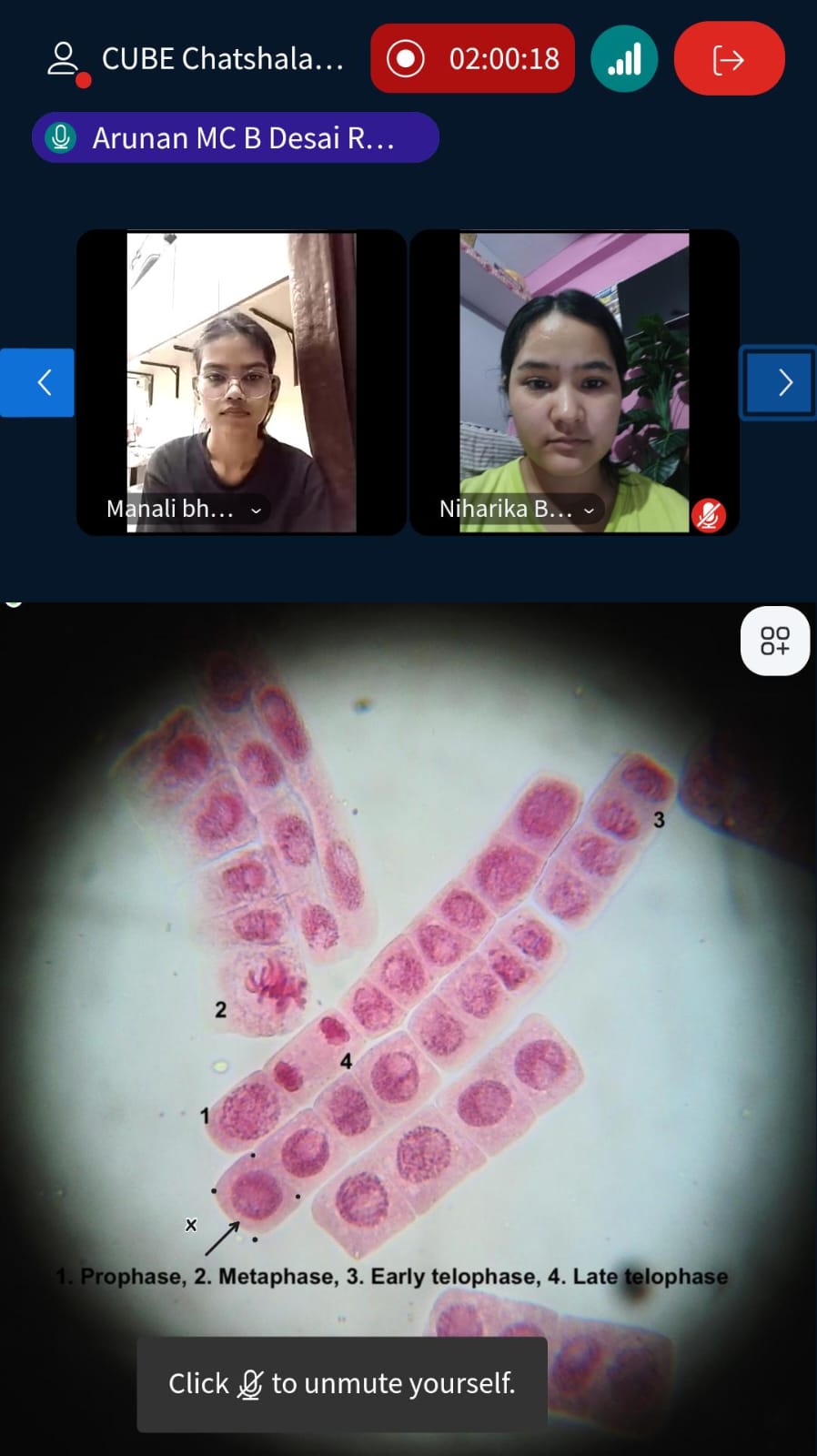
 Referance