CUBE ChatShaala — Discussion Summary
Date: 21 April 2026
Today’s CUBE ChatShaala session, held on 21 April 2026, revolved around an ongoing citizen science investigation into nail growth rates, focusing on young individuals in the 15–20 age group. The session brought together students and a mentor (Rechel mam) to present, compare, and critically examine data collected from real human subjects using a low-cost but methodologically sound marking technique.
The central research question driving the session was: How much do nails grow in 10 days in female and male subjects of the 15–20 age group? Two primary contributors — Niharika and Rechel — presented their hypotheses and supporting data, which opened up a rich discussion on both the biological and methodological dimensions of the study.
The Marking Methodology
The technique used across all subjects was a nail-marking method where a thin line is drawn on the nail surface at a fixed reference point (the proximal end of the visible nail) on Day 0. As the nail grows distally, the mark moves forward, and the distance between the original mark and the cuticle line gives a measurable estimate of nail growth. This is a clever, non-invasive way to observe nail growth without any special laboratory equipment.
Photographs were taken against graph paper to allow measurement. The images shared during the session included:
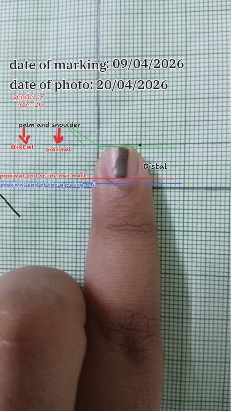
A female subject, age 20 (Gender: F), with a marking date of 09/04/2026 and a photo date of 20/04/2026. The annotated image carefully distinguished between the distal and proximal ends of the nail, and labelled the proximal end of the nail mark (red line) and the proximal end of the growing nail (blue line). The green lines indicated the palm-and-shoulder (distal) and proximal reference landmarks.
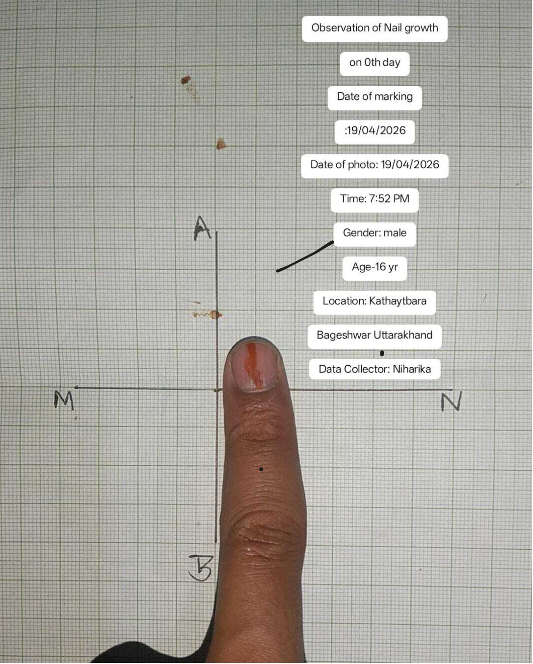
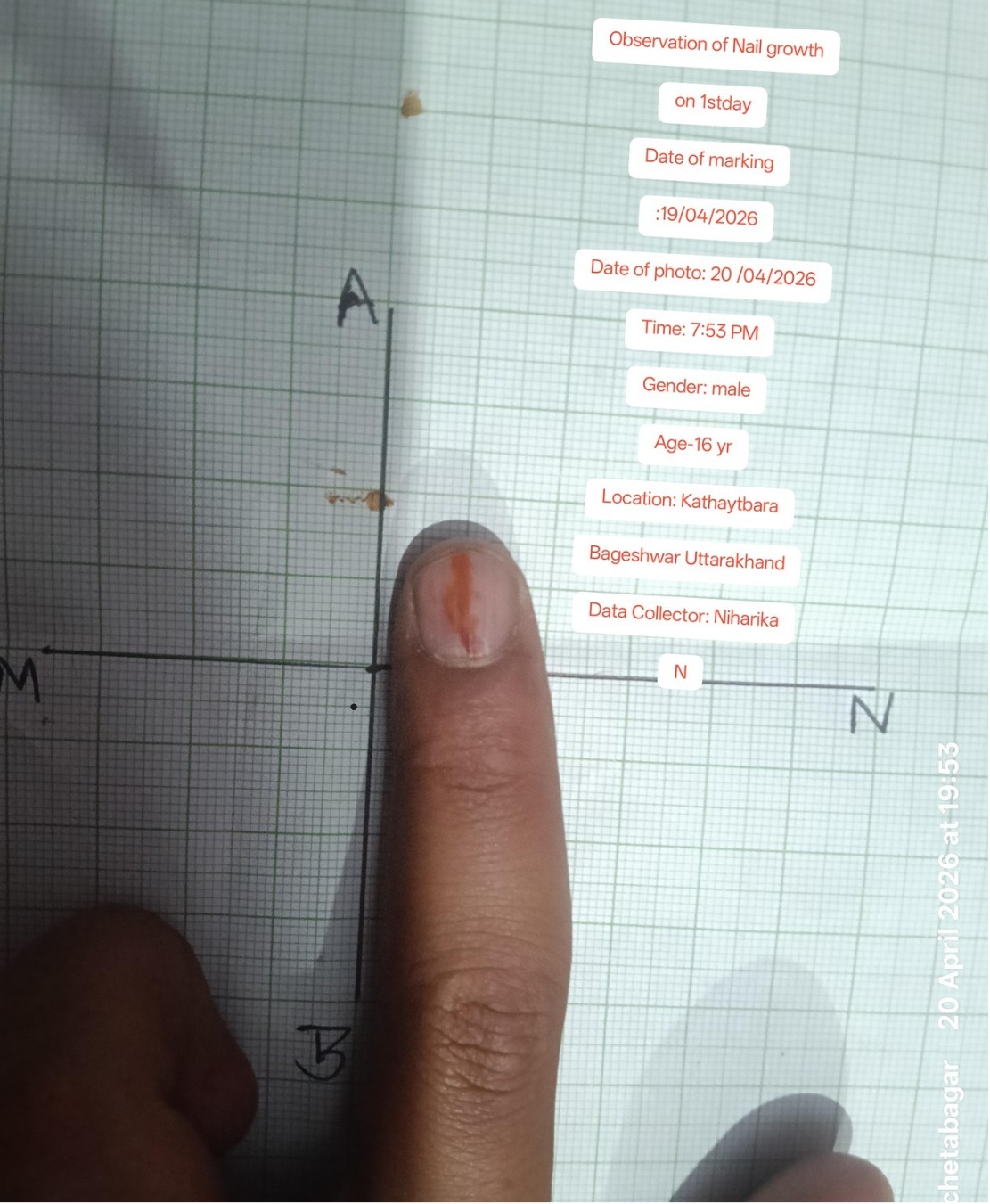
A male subject, age 16 (Data Collector: Niharika), from Kathaytbara, Bageshwar, Uttarakhand. Two photos were provided — one on Day 0 (marking date and photo date: 19/04/2026, Time: 7:52 PM) and one on Day 1 (photo date: 20/04/2026, Time: 7:53 PM). The finger was photographed against graph paper with coordinate axes labelled A–B (vertical) and M–N (horizontal), with a red nail mark clearly visible.
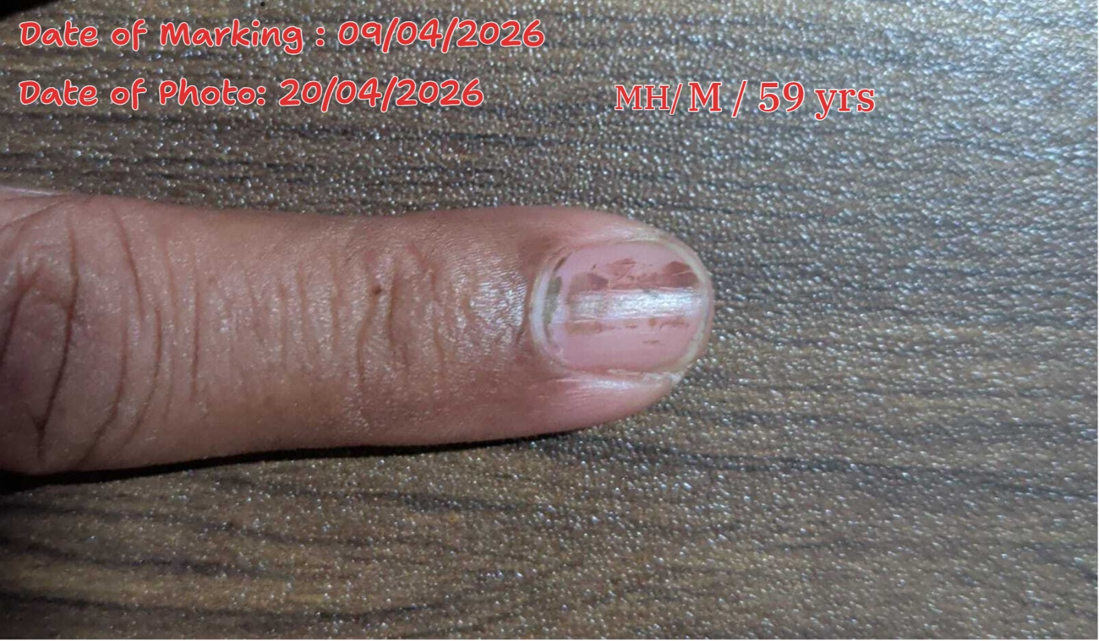
A third subject — an adult male from Maharashtra, aged 59 years (MH/M/59 yrs) — had a marking date of 09/04/2026 and a photo date of 20/04/2026. This subject introduces an interesting cross-age variable, extending the dataset significantly beyond the 15–20 target group. The nail appeared noticeably different in texture and had a thicker, more ridged appearance compared to the younger subjects.
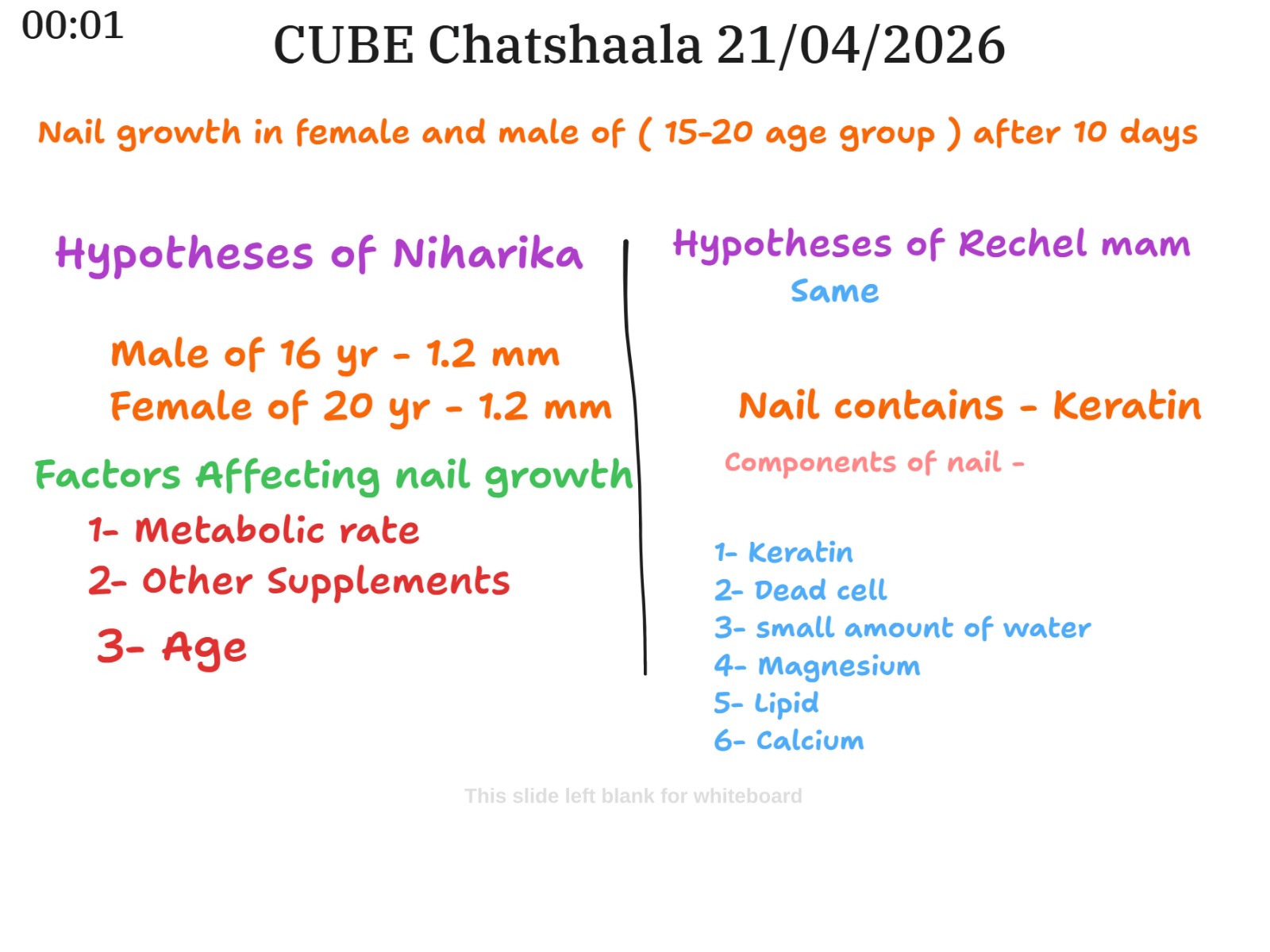
Hypotheses Presented
Niharika’s hypothesis predicted that both the 16-year-old male and the 20-year-old female would show a growth of approximately 1.2 mm over 10 days. Rechel mam’s hypothesis was noted as “same,” suggesting agreement with Niharika’s projection. Both hypotheses align loosely with scientific literature, which indicates that fingernails grow at roughly 3 mm per month on average, which would correspond to about 1 mm per 10 days.
Factors Affecting Nail Growth — Discussed in the Session
The whiteboard from the session listed three major factors that participants identified as potentially influencing nail growth:
Metabolic rate — a higher metabolic rate may correlate with faster cell division in the nail matrix, which drives growth. Other supplements — nutritional inputs such as vitamins and minerals may support or limit growth. Age — widely considered one of the strongest biological determinants of nail growth rate.
Nail Composition — Discussed in the Session
An important part of the session involved Rachel’s mom elaborating on what nails are actually made of. The whiteboard listed the following components: keratin, dead cells, a small amount of water, magnesium, lipids, and calcium.
This is largely consistent with scientific literature. The nail plate is predominantly composed of hard alpha-keratin, a fibrous scleroprotein that forms through tightly packed, layered dead keratinocytes. Scientific research confirms that the nail plate consists of roughly 80–90% hard alpha-keratin, with the remainder comprising soft epithelial keratins. Small quantities of water, lipids (0.1–1%), and various mineral elements, including calcium, magnesium, iron, zinc, phosphorus, and sulfur, are also present. Notably, calcium is found in the nail but is not the structural backbone — that role belongs entirely to keratin, which means the common belief that nails are made of calcium is a misconception worth addressing.
 Provocative Questions
-
The two hypotheses (Niharika’s and Rechel mam’s) both predict 1.2 mm growth in 10 days for subjects of different ages and genders. If the actual data support this prediction for both, does that mean age and gender do not significantly affect nail growth in the 15–20 range, or could the sample size be too small to reveal real differences?
-
The 59-year-old male subject was included in the dataset alongside 15–20-year-olds. Given that scientific literature consistently reports slower nail growth with advancing age, how might including this data point skew the group’s conclusions? Should older subjects be analyzed in a separate cohort entirely?
-
The marking technique relies on photographing the nail against graph paper. What are the potential sources of error in this method? For instance, could the angle of photography, the width of the pen mark, or the slight movement of the finger between photos affect the accuracy of the measurement?
-
The session noted metabolic rate as a factor affecting nail growth. How would you design a controlled experiment to test whether a higher metabolic rate (say, in a subject who exercises heavily) actually corresponds to faster nail growth compared to a more sedentary individual of the same age and gender?
-
Nail composition was listed on the whiteboard as including keratin, dead cells, water, magnesium, lipid, and calcium. But the dominant structural protein is alpha-keratin. If calcium is a minor component, why do so many people — and sometimes even students of biology — believe nails are made primarily of calcium? Where does this misconception come from, and how can it be addressed?
-
The Day 0 and Day 1 photographs for the 16-year-old male show very little visible change. Is it realistic to expect measurable growth within a single day? What is the minimum time interval that would give a reliably measurable result using the graph paper method?
-
Two subjects shared the same marking date (09/04/2026), but their photos were taken on 20/04/2026, giving an 11-day interval rather than exactly 10. How important is it to be precise about the number of days when calculating a daily or 10-day growth rate? Would even a 1-day difference in the interval significantly affect the final calculation?
 What I Have Learned
The first and most striking realization was how elegant and accessible the nail-marking method really is. It requires no laboratory equipment — just a pen, graph paper, a ruler, and a camera. Yet when done carefully with documented dates, times, subject metadata (age, gender, location, data collector), and a clear reference grid, it becomes a genuinely rigorous scientific protocol. This is the essence of CUBE’s approach: turning everyday biology into a site of real inquiry.
Second, we learned to think more carefully about variables. Age was listed as a factor affecting nail growth, and this makes biological sense — younger individuals tend to have faster cell turnover in the nail matrix. But the session also showed that variables like metabolic rate and nutrition are harder to control in a citizen science setting. This is not a flaw; it is actually what makes the data richer and more interesting when pooled across many contributors.
Third, the composition of nails came into much clearer focus for me. Before today, probably would have repeated the common assumption that nails are mainly calcium. But they are not — they are predominantly hard alpha-keratin, a fibrous structural protein that forms tight intermediate filaments. Calcium, magnesium, and lipids are present but play supporting roles. This distinction matters because it changes how we think about nail health and what dietary factors actually support nail strength.
Finally, we were reminded of the importance of documenting everything — subject details, exact dates and times, location, and data collector name. The photographs shared today were excellent examples of this practice. Each image functioned not just as a picture, but as a data point with a full metadata label. That level of care is what separates anecdote from evidence.
 TINKE Moments ("This I Never Knew Earlier)
TINKE 1: Nails are not made of calcium.
The whiteboard listed calcium as one component of nail structure, which it is — but as a minor mineral element, not a structural one. The primary material is alpha-keratin. Many participants likely held the folk belief that strong nails = calcium, the same way bones = calcium. This session planted the seed of doubt about that assumption, and following up on the actual science reveals a very different story. The keratin-dominant structure of nails is a significant TINKE.
TINKE 2: The nail mark moves — the nail doesn’t just get longer at the tip.
Students sometimes assume that nail growth means the tip of the nail gets longer, and that is where one should look for growth. But the marking technique demonstrates that growth occurs proximally, from the nail matrix, and the entire nail plate moves distally as a unit. The mark placed near the cuticle moves toward the free edge. This spatial understanding of nail growth direction is a genuine conceptual shift.
TINKE 3: Day 0 and Day 1 photos show almost no visible change.
Several participants may have expected to see a noticeable difference even within 24 hours. Seeing that the Day 1 photograph of the 16-year-old male showed barely any change compared to Day 0 is a reminder that biological processes unfold at their own pace — and that impatience in measurement leads to uninterpretable data.
TINKE 4: Older nails look structurally different — even in photographs.
The photograph of the 59-year-old male subject showed a visibly different nail compared to the younger subjects — thicker, with longitudinal ridges and a more opaque appearance. This is a natural result of the ageing of the nail matrix and changes in keratin turnover. Many participants may not have connected the visible appearance of an elderly person’s nails to the underlying cellular biology. Seeing it in a real photograph makes this connection tangible.
 Gaps and Misconceptions
Gap 1 — Sample size and generalizability.
The current dataset appears to involve only a handful of subjects. Drawing conclusions about gender- or age-based differences in nail growth from two or three individuals is statistically premature. A larger, more diverse sample — with the same gender, age, location, and hand dominance documented for every subject — would be needed before any hypothesis can be confirmed or refuted.
Gap 2 — No mention of which finger was used.
Nail growth rates differ between fingers. Scientific literature notes that the nail of the index finger grows faster than that of the little finger, and the dominant hand tends to show slightly faster growth than the non-dominant one. None of the photographs or whiteboard notes mentioned which finger was being photographed. This is a variable that should be standardized or at least documented.
Gap 3 — Measurement technique not yet quantified.
The session showed photographs against graph paper, but the actual measurements in millimetres were not discussed in detail beyond the hypothesized values of 1.2 mm. It would strengthen the project considerably to show how participants measured the displacement of the mark from the original position and calculated the actual growth in mm from the graph paper grid.
Misconception 1 — Calcium builds nails.
As discussed above, this is a widely held but scientifically inaccurate belief. Nails are built from keratin, not calcium. While mineral deficiencies can affect nail health, supplementing calcium will not necessarily improve nail strength the way improving protein (specifically keratin precursor amino acids like cysteine and methionine) would.
Misconception 2 — Nail growth can be measured within a day.
Including a Day 0 to Day 1 photograph pair may inadvertently suggest to newer participants that daily tracking is meaningful. In reality, with an average growth of approximately 0.1 mm per day, the graph paper method will not reliably resolve such small differences. A 10-day or longer interval is much more appropriate for this protocol.
Misconception 3 — Gender alone determines growth rate.
The hypothesis as framed compares a 16-year-old male with a 20-year-old female. These subjects differ in both age and gender simultaneously, making it impossible to attribute any difference in growth solely to gender. To isolate gender as a variable, the comparison should be made between subjects of the same age and different genders, or vice versa.