AFTER 3 DAYS
 Male Belly-button
AFTER 3 DAYS
Male Belly-button
AFTER 3 DAYS
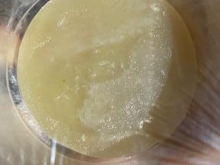 Female ear
AFTER 3 DAYS
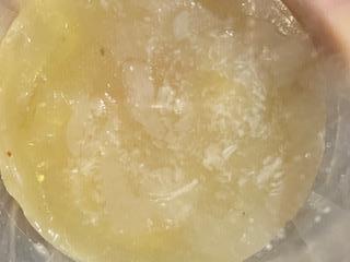
 Male ear
Good that you’ve been following up with photo evidence. But, to a viewer, all the observations are appearing to be nonetheless the same due to the texture of the media and also, the way by which the photograph has been taken.
To make things clear, can you elaborate on the observations that you’ve got?
Any colonies? If yes, then what is their colour, texture?
Something like colony characteristics, if any?
Good luck!
Below is the video we used to make the homemade petri dishes:

Instead of using the beef bouillon cube, we used chicken stock.
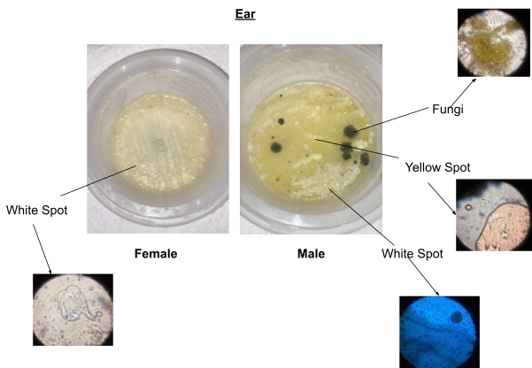
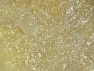
In the female ear, we can spot white spots, which seem to be bacteria, once looked through the foldscope. However, in the male ear, not only are there white spots but also fungi and yellow spots. This was very interesting to see!
Female ear: White Spots
Male ear: White Spots, Fungi, Yellow Spots
Experiment held: 25/08/21 - 27/08/21
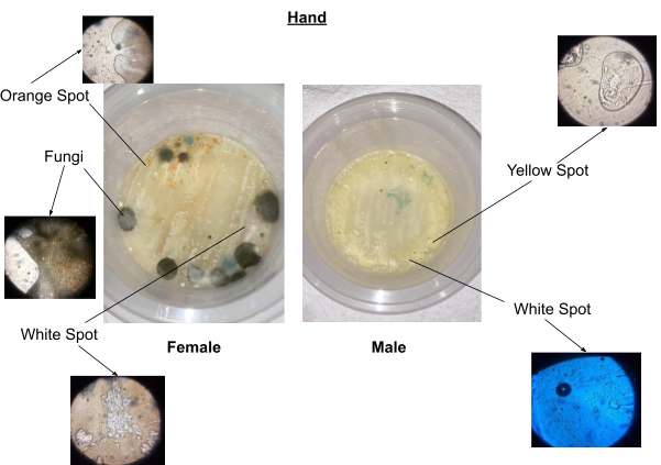
On the female hand, we can see many species of what seem to be bacteria; orange spots and white spots, and also fungi. However, on the male hand, there seems to be less growth and only two species: yellow spot and white spot.
Female Hand: White Spot, Fungi, Orange Spot
Male Hand: White Spot, Yellow Spot
Experiment held: 25/08/21 - 27/08/21

In the female belly button, we can see that there is a lot of fungi growth on one half and slight growth of orange spots on the other. However, in the male belly button, there is a new species that is pink in colour. This was very fascinating!
Female Bellybutton: White Spot, Fungi, Orange Spot
Male Bellybutton: White Spot, Yellow Spot, Pink Spot
Experiment held: 25/08/21 - 27/08/21
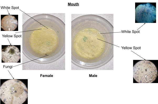
The female mouth had a little bit of fungi on the side of the container.
Female Hand: White Spot, Fungi, Yellow Spot
Male Hand: White Spot, Yellow Spot
Experiment held: 25/08/21 - 27/08/21