 CUBE ChatShaala — Discussion Summary
Date: 26 April 2026
Today’s CUBE ChatShaala session centred on something deceptively simple — the fingernail. What started as an observation about a red streak visible beneath a nail quickly opened into a wide-ranging conversation about nail anatomy, the sensory biology of the fingertip, and what it really means to “observe” in science.
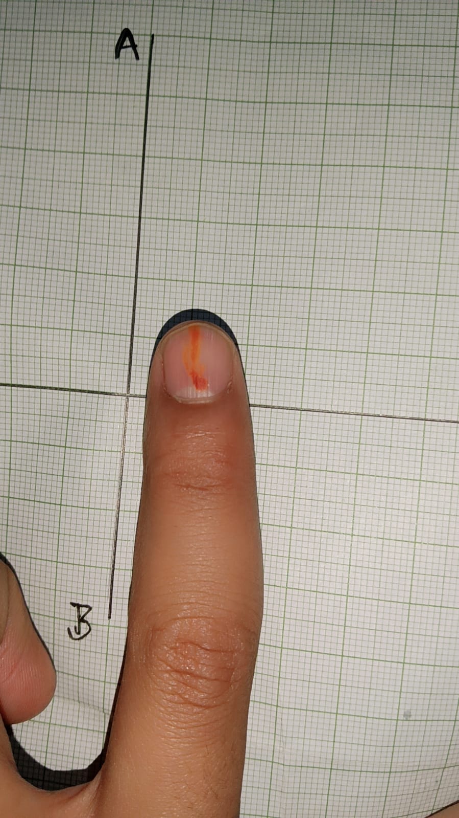
The session opened with a participant sharing an image (dated 25 April 2026) of their own fingertip, held against graph paper marked with axes A and B. Clearly visible beneath the nail was a thin, bright orange-red henna longitudinal streak running from near the cuticle toward the free edge. This was not just a visual curiosity — it became the anchor for the entire discussion.
Key observation: A participant noticed a reddish streak running lengthwise under the nail — possibly a nutritional deficiency. The question posed: * Why does discoloration under the nail appear as a narrow longitudinal streak instead of spreading out diffusely?*
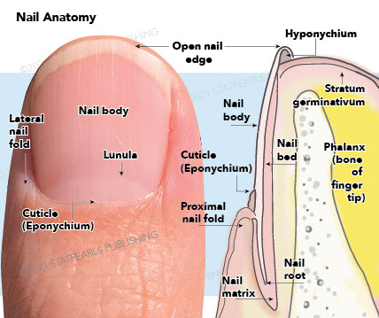
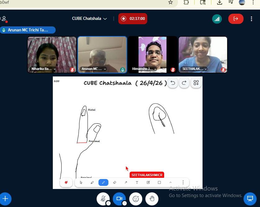
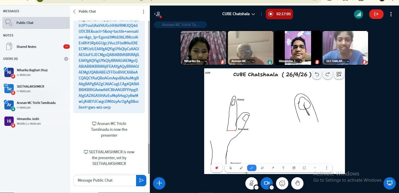
The group then worked to understand the structural anatomy of the nail — using both a labelled diagram shared in the session and hand-drawn sketches on the shared whiteboard. Participants drew and discussed the orientation of the finger, correctly labelling the distal (tip) and proximal (base) ends. A second sketch showed the lateral profile of the finger with the nail root visible beneath the skin fold.
Discussion covered the following anatomical structures:
- Nail body — the visible hard plate
- Nail matrix — the growth zone, located proximally beneath the skin; damage here affects nail shape permanently
- Nail bed — the pink vascular tissue beneath the nail plate, responsible for nourishment and attachment
- Lunula — the pale crescent at the nail’s proximal end, the visible portion of the matrix
- Cuticle (Eponychium) — the thin skin sealing the proximal nail fold
- Hyponychium — the skin seal beneath the free edge of the nail
- Lateral nail fold — the skin flanking the nail on the sides
- Nail root — embedded portion, hidden under the skin
A key conceptual point that emerged: the nail bed has a highly organised, longitudinal vascular architecture. The blood vessels in the nail bed run in parallel channels from proximal to distal. When one of these small vessels leaks or ruptures (as in a splinter haemorrhage), the blood cannot spread laterally because it is constrained by the rigid nail plate above and the channelled bed below — producing that characteristic linear red streak.
The group also touched on the role of the fingertip in tactile sensation. The nail itself is not sensitive, but it acts as a rigid counter-pressure surface against the soft pulp of the fingertip, enhancing the ability to detect fine textures and pressure. Specialized receptors (Meissner’s corpuscles, Merkel discs) are densely packed in the fingertip skin, particularly under and around the nail bed area, making fingertips one of the most touch-sensitive regions of the human body.
The whiteboard sketch drew attention to the proximal-to-distal orientation — a recurring theme in biology, indicating direction of growth and structural polarity. The group discussed that nails grow from the matrix outward (distally) and that the nail plate slides over the nail bed as it elongates.
 Provocative Questions
-
The blood under the nail forms a streak, not a blob. What specific structural features of the nail bed force blood to travel in a linear path rather than pooling or spreading?
-
If the nail matrix is damaged — say, by a heavy impact — the nail may grow deformed permanently. What does this tell us about where “information” for nail shape is stored in the body?
-
The lunula is visible on some fingers but not others, and is more prominent on the thumb. Is this a difference in anatomy or simply a difference in how deep the matrix sits relative to the fold? How would you design an observation to test this?
-
Fingernails and toenails grow at different rates. What variables — vascular supply, mechanical load, temperature — might explain this, and how would you isolate each factor?
-
The nail acts as a mechanical backstop for the fingertip, improving tactile acuity. If you had no nails at all, in what specific tasks do you predict your sensitivity or grip ability would be most impaired? How would you measure this?
-
When drawing the finger on the whiteboard, a participant initially struggled to correctly identify which end was proximal and which was distal. Why might directional orientation in anatomy feel unintuitive — and what does this reveal about how we typically observe our own bodies?
-
The hyponychium seals the underside of the free nail edge to prevent pathogens from entering the nail bed. What would happen structurally and biologically if this seal were consistently broken — as it often is in nail-biters
 What I Have Learned
The most important lesson from today is that a single, seemingly minor observation — a red streak under a nail — can serve as the entry point for a rigorous investigation of anatomy, vascular biology, and sensory physiology. Science does not always begin with grand hypotheses; it often begins with noticing something small and refusing to dismiss it.
I learned that nail anatomy is far more layered than it appears. The nail is not just a hard cover — it is a complex unit involving the matrix (growth), the bed (support and vascular nourishment), the folds (protection), and the plate (mechanical function). Each structure has a distinct role, and disruption of any one part has cascading effects.
I also came to appreciate the concept of longitudinal vascular architecture in the nail bed. The reason a splinter haemorrhage is linear is not accidental — it reflects the underlying organisation of the tissue. Structure explains appearance. This is a principle that applies across all of biology.
On the pedagogical side, today reinforced the value of drawing and labelling as thinking tools. Sketching the finger on a whiteboard — even imperfectly — forces you to confront what you actually know versus what you merely assume you know. Several participants discovered their mental model of “proximal” and “distal” was less secure than they thought until they had to commit it to a diagram.
TINKE Moments (This I Never Knew Earlier )
TINKE 1 — The nail bed has directional vessels. Most participants assumed blood vessels in the nail bed were a general mesh or network. The realisation that they are longitudinally oriented — and that this explains why bleeds appear as streaks — was a genuine surprise. Prior assumption: vessels are randomly arranged. Corrected understanding: architecture is highly organised and functionally meaningful.
TINKE 2 — The nail root is hidden, not visible. When asked where the nail begins, several participants pointed to the cuticle line. The correction — that the nail root is embedded several millimetres beneath the proximal nail fold and is never visible — prompted visible surprise. The lunula is not the root; it is merely the edge of the matrix peeking through.
TINKE 3 — Proximal vs. distal confusion in self-observation. When drawing one’s own finger on the whiteboard, orienting which end is “proximal” (closer to the body) and which is “distal” (further from the body) caused hesitation. This revealed that anatomical directional terms, while learned, are not always intuitively applied to one’s own body — especially for a structure as familiar as a finger.
TINKE 4 — The nail enhances touch, not just protects. The common understanding is that nails protect the fingertip. The additional insight — that the nail’s rigidity provides counter-pressure that sharpens tactile discrimination — was new for several participants. You feel texture more precisely because the nail gives sensory receptors something solid to work against.
TINKE 5 — The hyponychium exists and matters. The seal of skin beneath the free nail edge (hyponychium) was unknown to most participants by name and by function. Its role in preventing microbial entry into the nail bed highlights how even “unremarkable” tissue has an important biological purpose.
 Gaps and Misconceptions
Gap 1 — Rate and control of nail growth. The group discussed growth direction but did not explore the regulatory biology: what controls the rate of nail growth? Are there hormonal, nutritional, or circadian factors? This remains an open question for future sessions.
Gap 2 — Clinical significance of splinter haemorrhages. The red streak was observed and explained structurally, but its clinical relevance (e.g., as a sign of infective endocarditis, vasculitis, or trauma) was not fully discussed. This is a meaningful gap — the same observation that started the session has significant diagnostic weight in medicine.
Gap 3 — Comparison across species. Do other primates have the same nail bed architecture? Do claws (in cats, dogs) work differently? The evolutionary and comparative dimension was not touched, which could have deepened the understanding of why the human nail has its particular structure.
Misconception — “The lunula is where the nail grows.” This is a common lay belief. The lunula is the visible portion of the nail matrix, but growth occurs throughout the matrix — not just at that crescent-shaped tip. This was partially addressed but warrants a clearer, more explicit correction in future sessions.
Misconception — “Cutting nails causes pain because of nerves in the nail.” The nail plate itself has no nerve supply. Any sensation during cutting comes from the surrounding soft tissue or, if cut too short, from the nail bed. This distinction between nail plate (insensate, keratinised) and nail bed (sensate, vascularised) is foundational and should be stated explicitly.