 CUBE ChatShaala Summary – 18.10.2025
Topic: Immunohistochemistry and Western Blot
Today’s CUBE ChatShaala focused on understanding two key molecular biology techniques—Immunohistochemistry (IHC) and Western Blotting, both essential in detecting specific proteins within cells and tissues.
 Key Discussion Points:
1. Immunohistochemistry (IHC):
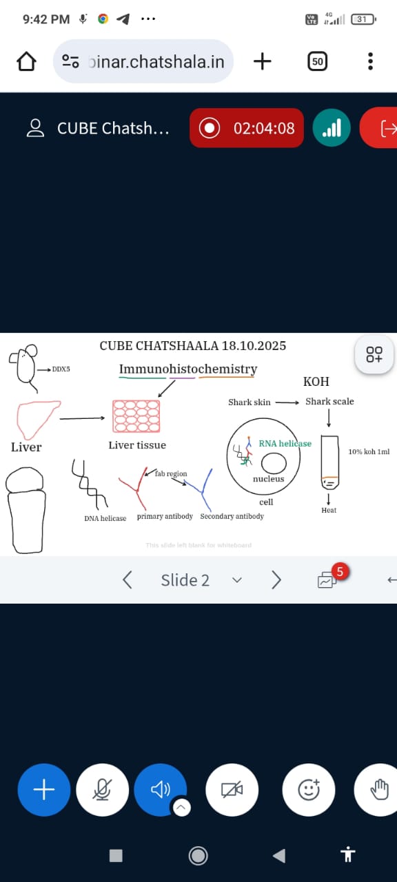
The session began by discussing the use of liver tissue as a model system to detect the protein DDX5, an RNA helicase involved in RNA metabolism.
-
The process involved treating tissue sections with primary antibodies that specifically bind to DDX5, followed by secondary antibodies linked to a reporter for visualization.
-
The diagram also indicated the Fab region of antibodies and the interaction between the nucleus, RNA helicase, and cellular localization.
-
A side discussion explored Shark scale preparation using KOH treatment (10%) and heating, possibly for studying tissue structure or antigen retrieval.
This integration of biological structure (like shark skin) with molecular detection methods sparked curiosity about comparative tissue analysis.
2. Western Blot:
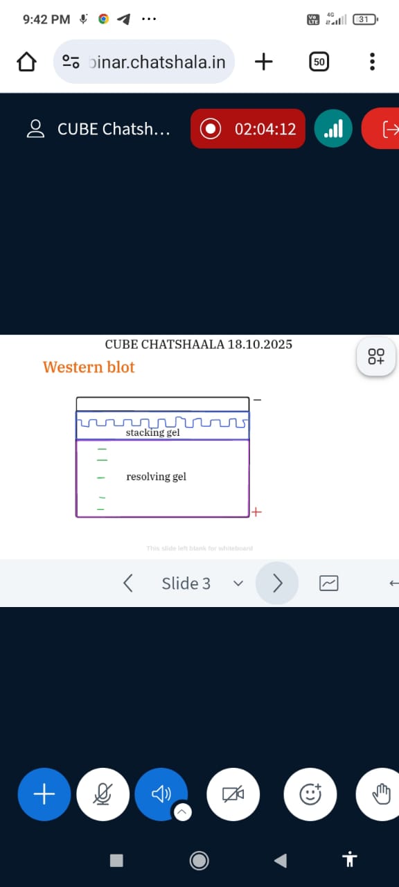
The second part of the meeting introduced the Western blot apparatus, emphasizing the two-layer gel structure:
-
Stacking gel – aligns proteins before separation.
-
Resolving gel – separates proteins based on molecular weight when an electric current is applied.
Participants connected this process to how it complements IHC — while IHC localizes proteins in tissues, Western blotting quantifies and verifies their presence.
 What I Learned:
-
I understood how antibody–antigen interactions form the foundation of both IHC and Western blotting.
-
The difference between localization (IHC) and quantification (Western blot) became clearer.
-
The session highlighted the significance of RNA helicase DDX5 as a biological marker.
-
The creative link between shark skin scale preparation and histochemical processing broadened the discussion toward structural biology.
 TINKE Moments (This I Never Knew Earlier)
-
Realizing that heat and KOH treatment might mimic antigen retrieval methods used in immunohistochemistry.
-
The Fab region of the antibody was discussed not just structurally but as a crucial player in specificity—linking structure to function beautifully.
-
The idea of comparing shark tissue with mammalian liver tissue revealed a cross-species curiosity—an important CUBE hallmark of connecting classroom learning to natural observations.
 Gaps and Misconceptions Identified:
- Some confusion remained about how RNA helicase (a nuclear protein) can be detected in tissue sections without degradation—clarification needed on fixation methods.
- A few participants mixed up Western blot with SDS-PAGE, highlighting the need to revisit how blotting adds antibody-based detection after electrophoresis.
- Questions arose about whether KOH treatment affects antigenicity and how it differs from traditional antigen retrieval methods in IHC.
 Provocative Questions for the Community:
-
 How does the specificity of the antibody in IHC determine the accuracy of protein localization?
 -
 Can shark skin or scale tissues be used as an experimental model for immunohistochemistry?
 -
Why do we use both IHC and Western blot to study the same protein—what unique insights does each provide?
-
 Does KOH treatment alter protein structure in a way that mimics biological denaturation, or can it preserve antigen sites?
 -
What are the creative ways we can visualize protein interactions in non-model organisms using simple materials?
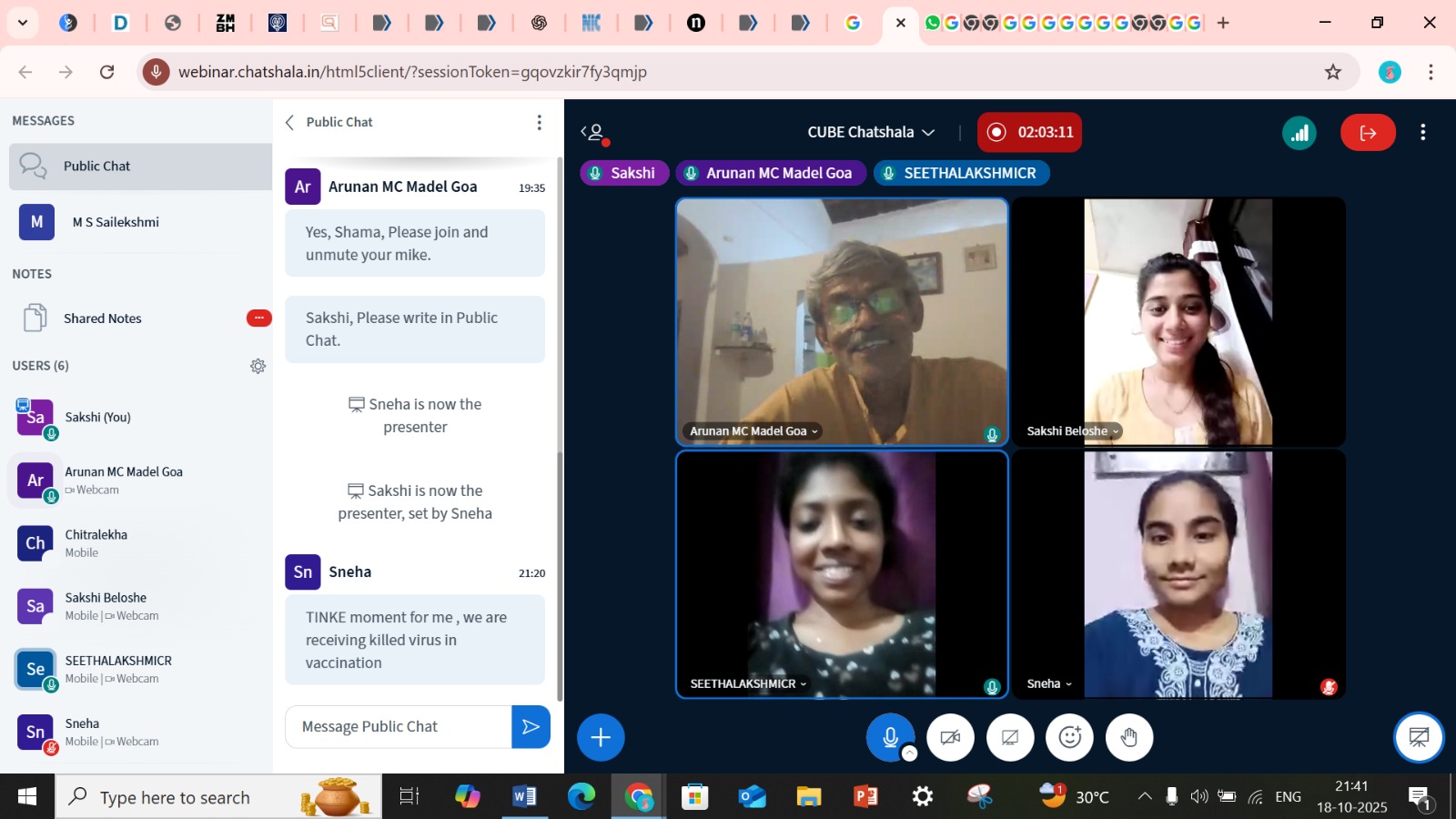
 photographs during Chatshaala
 Reference
-
https://www.nature.com/articles/s41401-020-00539-7?utm_source=chatgpt.com
-
https://www.sciencedirect.com/science/article/abs/pii/S0740257024000467?utm_source=chatgpt.com
-
🍀 When Antibodies Meet Colors: The Art and Science of Immunohistochemistry
@Arunan @2020ugchsncnseethala @SN1261 @Chitralekha and others.