 CUBE ChatShaala Summary – 17 October 2025
Today’s CUBE ChatShaala delved into the fascinating world of Immunohistochemistry (IHC) — a method used to detect specific antigens in biological tissues using antibodies. The discussion began by differentiating between Cryostat and Microtome, both instruments used for cutting tissue sections, and moved on to understand their relevance in IHC techniques.
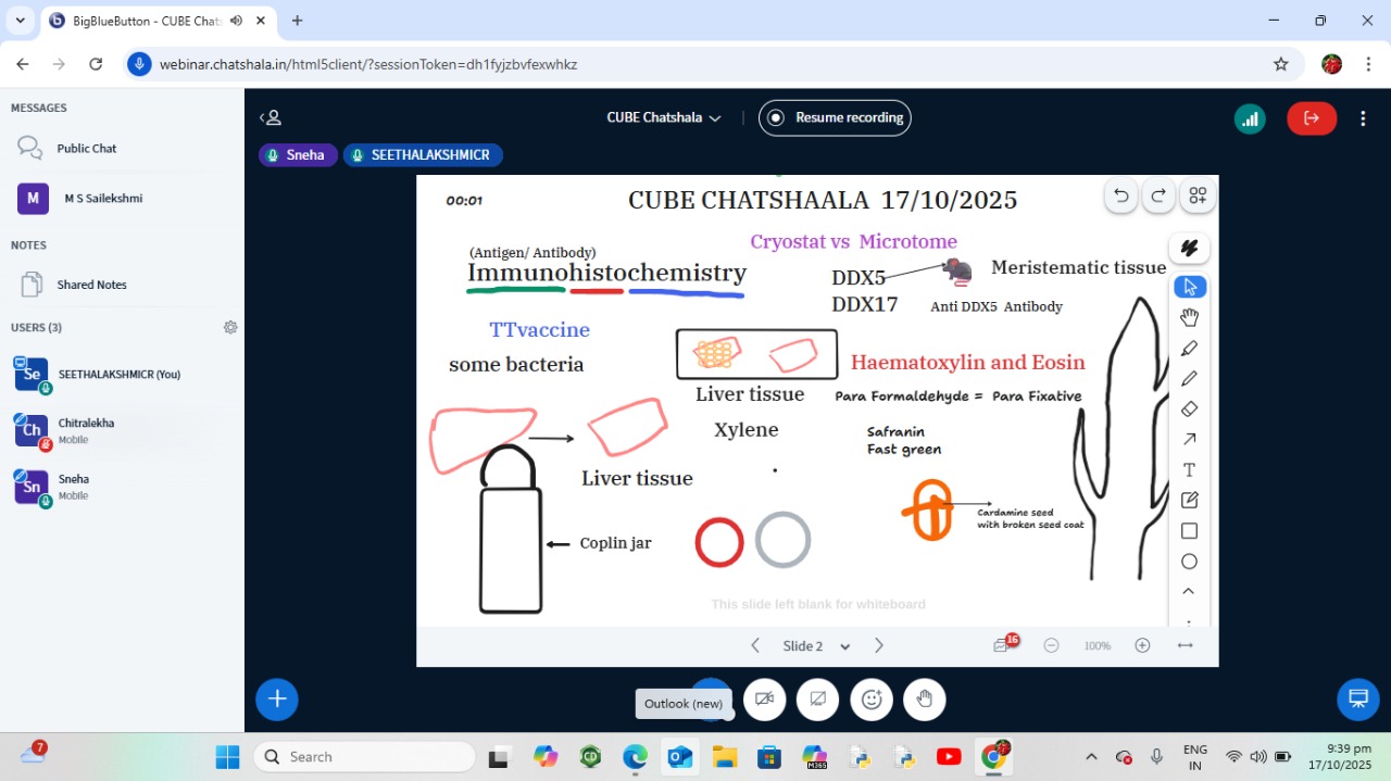
Participants discussed liver tissue preparation and the Coplin jar method, emphasizing the importance of xylene for tissue clearing. The use of formaldehyde (paraformaldehyde) as a fixative was explained, highlighting how it preserves cellular architecture. The staining techniques using Haematoxylin and Eosin (H&E), as well as Safranin and Fast Green, were explored to understand tissue visualization under a microscope.
A major highlight was the mention of DDX5 and DDX17, RNA helicases studied using anti-DDX5 antibodies, showing the biological applications of IHC in identifying specific proteins. The discussion also touched upon the TT vaccine and the role of antigens and antibodies in immune responses, drawing parallels between immunology and histology.
The session concluded with observations of Cardamine seeds with broken seed coats, connecting the structural biology of plant tissues (like meristematic tissue) to the techniques used for animal tissue studies. This blend of plant and animal biology sparked curiosity about how similar principles of fixation and staining can apply across biological systems.
 TINKE Moments (This I Never Knew Earlier)
-
The realization that formaldehyde acts as a “para-fixative,” preserving both tissue structure and antigenicity for IHC, was a key insight.
-
The visual link between Cardamine seed coat breakage and tissue sectioning techniques deepened the understanding of how sample preparation influences microscopic analysis.
-
The discussion connected TT vaccine’s antigen-antibody reaction with IHC principles, bridging immunology and histology in a thought-provoking way.
 Gaps and Misconceptions Identified
-
Confusion between Cryostat and Microtome use: Cryostat is for frozen sections, while
Microtome is for paraffin-embedded tissues. -
Some uncertainty remained about why xylene is specifically used during tissue processing.
-
The difference between Haematoxylin (nuclear stain) and Eosin (cytoplasmic stain) needed more clarity during the session.
 Provocative Queries for the Community
-
Can a plant tissue like Cardamine meristem be stained using the same H&E technique used for liver tissue?
-
How does the fixation process in IHC ensure that antigens remain detectable even after chemical treatment?
-
Can immunohistochemistry be extended to visualize bacterial proteins, like those in TT vaccine preparation?
-
If Cryostat preserves antigenicity better, should it replace Microtome in all IHC studies?
 What I Learned
Today’s ChatShaala beautifully bridged the worlds of immunology, histology, and plant biology. I learned that IHC is not merely a staining process—it’s a delicate combination of chemistry, biology, and microscopy. Understanding how fixatives, stains, and antibodies work together deepens appreciation for how microscopic observations can reveal molecular-level phenomena.
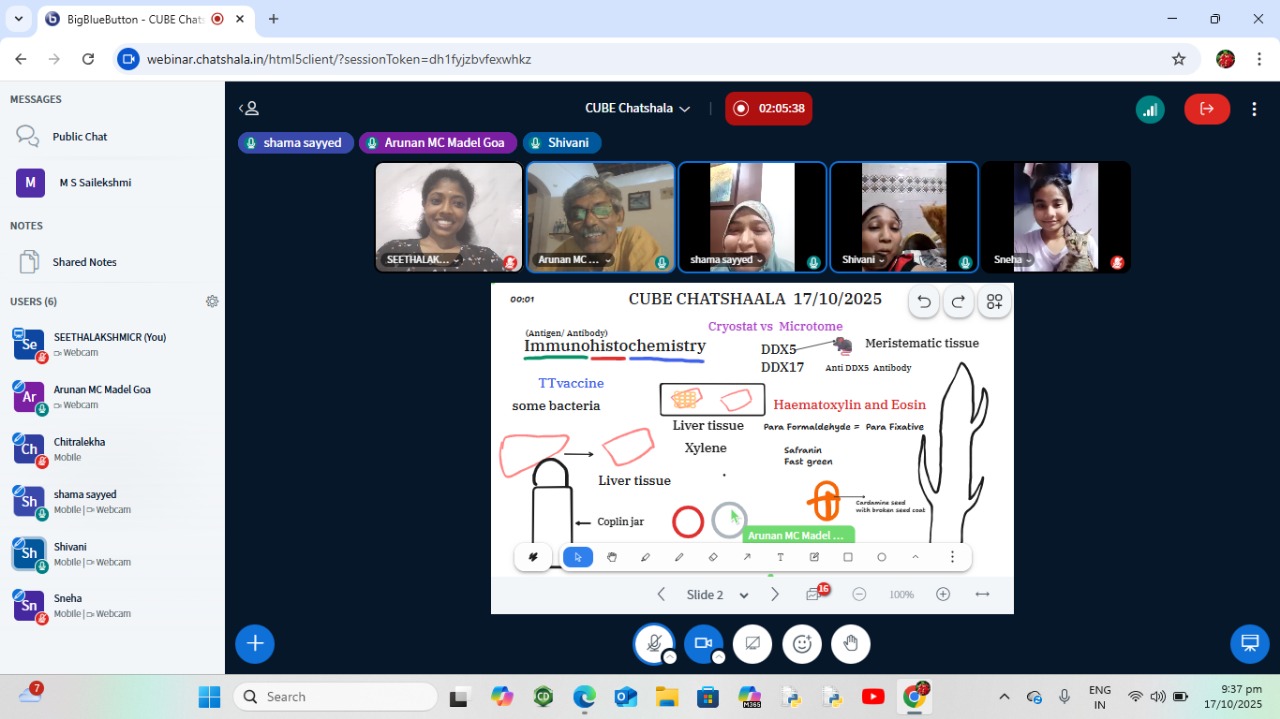
 photographs during Chatshaala

 Reference
-
Histological-Based Stainings using Free-Floating Tissue Sections - PMC
-
https://www.sciencedirect.com/topics/neuroscience/cryostat?utm_source=chatgpt.com
@sakshiconsultant2002 @Arunan @SN1261 @Chitralekha @2020ugchsncnseethala and others